

Why is understanding the anatomy of the skull more important than ever in 2025? As medical education and research evolve, choosing the right skull model for both study and clinical practice becomes crucial.
This guide helps you navigate the selection, use and understanding of skull models across medicine, education and research. You will gain insight into what a skull model is, the different types and materials, applications, selection criteria, maintenance and future trends.
Do you want to reap the benefits of proper selection and use? Read on and become an expert in skull models by 2025.
What is a skull model?
A skull model is an indispensable tool when it comes to understanding human and animal anatomy in depth. This section delves into what a skull model is, how it has evolved over time, and why it plays a central role in today's teaching and healthcare practice. Let's explore how the skull model differs across purposes, materials, and users.
Definition and historical development
A skull model is a physical or digital representation of the skull, used to illustrate and study the complex structures that make up the skeleton of the head. The purpose of a skull model has always been to facilitate the understanding of the details of anatomy, whether for teaching, research or clinical practice.
Historically, the development of the skull model has been characterized by innovation. In ancient times, models were created in wax or clay, often with limited accuracy. Later, plaster and resin came along, allowing for more detailed reproductions. Today, 3D printing has become a game changer, where precision and individualization are paramount. You can read more about this development in the Guide to Anatomical Models , which covers both the history and modern uses of the skull model.
Accuracy and realism have always been key aspects, as even small errors can lead to misunderstandings in anatomy teaching. Therefore, the quality of a skull model is more crucial than ever.
Anatomical details and structure
A skull model typically shows the main structures of the skull, such as the joints of the bones (sutures), openings (foramina), and the shape and thickness of the individual bones. The level of detail varies from simple models that only show the main structure, to advanced versions with separate bones and color-coded areas.
For example, an advanced skull model may have numbered markings for each bone, visible nerves and blood vessels, and realistically rendered bone surfaces, making it easier for users to identify and remember the various anatomical elements.
A simple model may be sufficient in basic teaching, but for specialized purposes such as surgical training or research, the most detailed skull model is often required.
| Model type | Level of detail | Typical users |
|---|---|---|
| Simple | Low | School, basic education |
| Advanced | High | University, clinic |
Differences between human and animal skull models
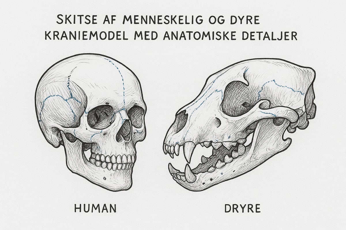
Both human and animal skull models are available, and the choice depends on the professional need. Human models are primarily used in medicine, dental education, and anatomy teaching, while animal models are often central to veterinary medicine and biological studies.
For example, an animal skull model can help understand the differences in dentition, jaw structure, and braincase shape between species, allowing for comparisons of evolutionary adaptations and biomechanical functions.
The use of both human and animal models provides a broader perspective and makes it easier to illustrate similarities and differences, which is crucial in both teaching and research.
Relevance in different fields of study
Today, the skull model plays a central role in many professional contexts. Medical students often use skull models to learn about the anatomy of the head, while dental students focus on the location of the dentition and its relationship to the rest of the skull.
In physiotherapy, understanding the structure of the skull is important for treating head and neck injuries. Biology students use skull models to investigate evolutionary relationships between species.
Statistics show that over 80 percent of medical students use physical models in their teaching, which emphasizes the importance of the skull model as a pedagogical tool.
The role of the skull model in modern anatomy teaching
In an age of digital alternatives, the physical skull model continues to play an important role in anatomy teaching. The tactile experience and the ability to examine the model from all angles enhances both understanding and memory.
Studies show that physical interaction with a skull model leads to better learning compared to using digital solutions alone. Many educators therefore combine physical models with apps and virtual tools to achieve the best learning outcomes.
The skull model is therefore not just a static object, but a dynamic tool that is constantly being developed to meet new demands in teaching and practice.
Types of skull models and materials
Choosing the right skull model requires insight into the different types and materials available on the market. Each model has unique characteristics, and it is crucial to match the need with the right solution for optimal learning and application.
Classic skull models vs. advanced models
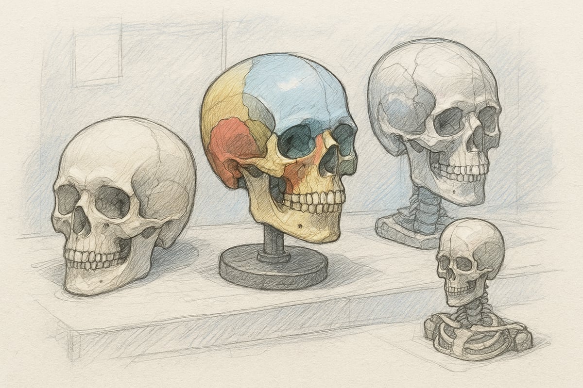
A classic skull model shows the basic anatomical structures and is often used for basic teaching. Advanced models can include details such as pathological changes, removable sections, or even built-in sensors for interactive learning.
Typical usage scenarios:
- Standard Models: Introduction to Anatomy in High Schools and Universities.
- Advanced models: Surgical training, specialized research, or specific disease studies.
The choice between a simple and advanced skull model depends on the desired depth of teaching and the user's professional needs.
Materials: Plastic, resin, 3D printing and natural materials
Skull models are made from several materials, each with its own strengths and weaknesses. Plastic is lightweight, durable and cost-effective. Resin provides high levels of detail and a more realistic feel. 3D printing allows for customized solutions and rapid production, while natural materials, such as real bone, are used in special cases.
| Material | Advantages | Disadvantages |
|---|---|---|
| Plastic | Cheap, light, robust | Less realistic |
| Resin | High detail, lifelike | More expensive, heavier |
| 3D printing | Adaptation, innovation | Can be fragile |
| Natural bone | Authenticity | Ethical/practical requirements |
When choosing a skull model, the material should match the purpose and expected wear.
Functional models: Movement and dismountability
Functional skull models are distinguished by their ability to be taken apart and show complex movements in the skull's joints, making them ideal for teaching where understanding spatial relationships and movement possibilities is central.
Examples of functionality:
- Removable skull caps or jaws.
- Movement in the jaw joint to illustrate chewing function.
- Magnetic or click systems for easy assembly.
A functional skull model increases interactivity and allows for hands-on learning, especially in clinical and educational contexts.
Color-coded and numbered models
Color coding and numbering make it easier to identify structures and remember complex anatomical relationships. Many students prefer a skull model with clear colors as it improves learning and overview.
Statistics show that 65% of students choose color-coded models to optimize their learning. For further guidance, visit For Anatomy Students , which offers resources for the effective use of skull models in teaching.
This type of skull model is especially popular in basic education and courses where visual differentiation is key to success.
Specialized skull models: Children, pathology and surgical training
Specialized skull models are developed for specific purposes, such as pediatrics, pathology, or advanced surgical practice. Models of pediatric skulls illustrate growth zones and development, while pathological models show disease changes such as fractures or tumors.
Benefits for specialized professional groups:
- Training in rare or complex cases.
- Preparation for surgical procedures.
- Focus on age-specific anatomy or disease patterns.
A specialized skull model provides unique opportunities for targeted training and research.
Price levels and availability
The price of a skull model varies considerably depending on the type, material and level of detail. Standard models can be obtained from around 500 DKK, while advanced or specialized models can cost several thousand DKK.
Where can I buy quality models?
- Professional suppliers of medical equipment.
- Specialty stores for educational materials.
- Online platforms with certified quality.
It is important to balance price and quality so that the skull model meets both budget and professional requirements.
Use of skull models in practice
Skull models have gained a central place in both teaching, clinical practice and research. As the demands for understanding anatomy increase, the use of skull models has become indispensable in many contexts. Here we review the most important practical applications that show why skull models are so essential today.
Teaching and learning
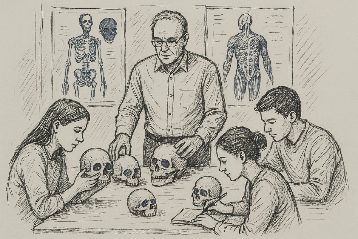
In educational settings, skull models are an indispensable tool for both teachers and students. The model is used in everything from elementary school to university level, where it creates a basis for practical learning and understanding of the complex structures of the skull. Many educators supplement physical models with digital resources and posters to enhance learning.
Teaching methods vary, but hands-on exercises with skull models promote both memory and spatial understanding. Students have the opportunity to examine sutures, foramina, and bone thickness, bringing the theory to life. For further inspiration, educators can find Tips for Teaching Anatomy , which provides concrete advice on integrating skull models into their teaching.
Clinical use: Doctors, dentists and therapists
In clinical practice, skull models are often used for patient communication and treatment planning. Doctors and dentists use the model to illustrate diseases, fractures or surgical procedures, helping patients understand their situation and treatment choices.
An example might be a surgeon using a skull model to explain an upcoming procedure to the patient. When the model is actively used in the conversation, the patient's confidence and understanding are increased. Therapists also use skull models in rehabilitation and retraining, where precise knowledge of anatomy is essential.
Research and development
Researchers use skull models in biomechanical testing and for the development of medical devices. The accuracy of a skull model is crucial when testing new implants or surgical techniques. 3D-printed models in particular have become popular since 2020, when their use has increased significantly.
Statistics show that more research environments are choosing advanced skull models to simulate realistic conditions. This enables testing of innovative solutions before they are used on patients. The precision requirements of research make choosing the right skull model an important factor in development work.
Simulation and skills training
Simulation training with skull models has revolutionized the education of surgeons and dentists. Realistic models allow for practice of procedures and skills before working on real patients. This increases both the safety and quality of treatment.
Examples of simulated procedures include drilling, cutting and assembling bone fragments. Using a skull model, students gain hands-on experience in a controlled environment. This improves both confidence and technical skills, which are essential for future healthcare professionals.
Public communication and museums
Skull models are also widely used in museums and for public education of anatomy. Physical model exhibits allow visitors to see and touch replicas of human skulls, creating engagement and learning across age groups.
Interactive learning environments that combine skull models with digital presentations provide an in-depth understanding of the structure of the skull. This communication strengthens interest in science and medicine, especially among young people and school classes.
Challenges and limitations
Although skull models have many advantages, they also have limitations. Physical models cannot always reproduce all anatomical variations or rare pathologies that digital and virtual solutions can simulate. Maintenance and storage also require resources.
Compared to digital alternatives, a physical skull model lacks the ability for rapid adaptation and interactivity. However, the tangible experience often provides better understanding and memory. The choice between physical and digital models therefore depends on purpose and resources.
How to choose the right skull model
Choosing the right skull model requires more than just choosing the first and best one. The process should start with a thorough analysis of what the model will be used for, what requirements are made for quality, materials and price, and where it can best be obtained. Here you will find a systematic guide so that you can make the best choice and get the most out of your investment.
Purpose and needs analysis
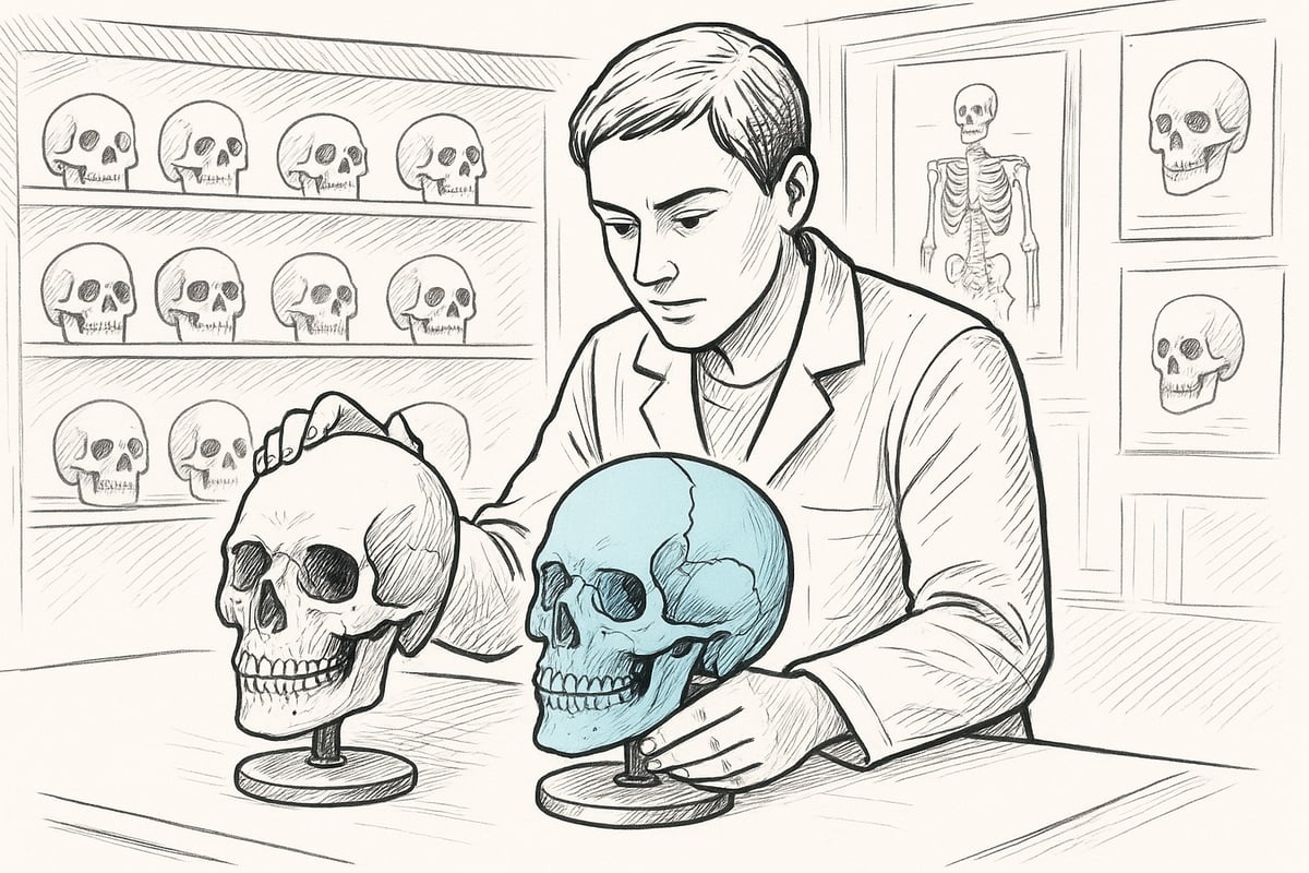
Before investing in a skull model, it is crucial to identify the specific need. Will the model be used for basic anatomy teaching, advanced surgical training, or clinical communication to patients? The purpose determines which features are most important.
Make a list of requirements, such as level of detail, ability to disassemble the model, or focus on specific age groups. Also consider whether there are any special requests for size or weight. By analyzing the purpose carefully, you ensure that the chosen skull model matches your expectations and usage scenarios.
Quality criteria and accuracy
The quality of a skull model is measured by its anatomical correctness and level of detail. A good model clearly shows sutures, foramina, and bone thickness. Also check if the model is based on authentic data or scans from real skulls.
Especially in teaching or research contexts, precision is essential. An accurate skull model increases understanding and minimizes the risk of misunderstanding. Always compare multiple models and note whether small anatomical variations are included to get the most value.
Material selection and durability
The material is important for both durability and realism. Plastic and resin are common choices as they are robust and easy to clean. 3D printed models allow for a high level of detail and customization, which is especially relevant for specialized needs.
If you are interested in how modern materials are used to create anatomical models, you can read about the Bachelor of Science in Industrial 3D Printing , which focuses on the design and production of, for example, skull models. Choose a material that suits your use so that your skull model lasts for many years.
Price vs. quality
Price and quality often go hand in hand when it comes to skull models. Budget models may be adequate for basic teaching, but typically lack detail and durability. Premium models are more expensive, but offer better realism, more features, and often a longer lifespan.
Consider whether it's worth investing a little extra to get a model that meets all your requirements and lasts longer. If necessary, make a comparison table of the options you're considering so you get an overview of what you're getting for your money.
Certifications and standards
When choosing a skull model for educational use, you should ensure that the model meets relevant certifications. CE marking is often a requirement in healthcare education and ensures that the model meets certain safety and quality standards.
Ask the supplier for documentation of certifications, especially if the model is to be used in exam situations or clinical teaching. A certified skull model is often a sign that the manufacturer is focused on both quality and user safety.
Purchase locations and suppliers
The final step in the process is to find a trustworthy supplier. Check if the supplier has good reviews and if they offer a warranty or the option to purchase spare parts. Be aware of copycat products, which are often of lower quality.
Look for specialty stores, reputable webshops, and dealers with experience in medical equipment. This way you will get the best service and ensure that your skull model meets all expectations and requirements.
Maintenance and storage of skull models
Proper maintenance and storage of your skull model not only ensures hygiene, but also that the model retains its function and details for many years. Whether you use your skull model in teaching, clinical practice or research, small routines can make a big difference to both durability and yield.
Cleaning and disinfection
Regular cleaning of your skull model is essential to maintain both hygiene and detail. Always start by brushing away loose dust with a soft brush. For daily wiping, a damp microfiber cloth is recommended that does not scratch the surface. For more thorough disinfection, you can use mild cleaning agents without alcohol, as strong chemicals can damage the material of the skull model. The frequency depends on use, but weekly cleaning is often sufficient in educational environments. Remember to let the model dry completely before storing it again.
Storage solutions
Storage plays a key role in the lifespan of a skull model. The model should always be stored in a dry room with a stable temperature and low humidity. Avoid direct sunlight, as UV rays can discolor and weaken the material. Use a dust-proof box or cabinet, preferably with padding, to avoid scratches and bumps. Many people choose to store their skull model together with related teaching materials such as books or anatomical posters and learning materials , which provides a clear learning environment. Regularly check the storage area for moisture and damage.
Repair and spare parts management
Even the most robust skull model can become worn or damaged over time. Minor damage, such as loose joints or small cracks, can often be repaired with special glue or replacement parts from the manufacturer. Many manufacturers offer replaceable parts, so you don't have to replace the entire model in the event of minor defects. It is important to document any repairs, especially if the model is used in an educational context. In the event of major damage, you should contact the supplier for professional assistance to ensure that your skull model remains functional and anatomically correct.
Long-term durability and warranty
To ensure the long-term durability of your skull model, you should follow the manufacturer's instructions for both use and storage. Most quality models have an expected lifespan of 5 to 10 years with proper maintenance. Many suppliers offer a warranty that covers defects and defects in the material, which provides extra peace of mind. Always keep your receipt and warranty certificate so that you can quickly get help if problems arise. By combining good maintenance with attention to the warranty, you can get maximum enjoyment from your skull model for many years.
Skull models of the future: Trends and innovations
The future of cranial modeling is marked by groundbreaking technologies and an increased focus on user experience, sustainability, and global accessibility. Cranial modeling is evolving rapidly, and new trends are making it possible to adapt models even more precisely for both teaching and clinical practice. Here, we review the most important innovations shaping the future of cranial modeling.
3D printing and digitization
3D printing has revolutionized the production of skull models. Today, precise copies of human skulls can be produced quickly and with great anatomical accuracy. This technology makes it possible to customize each skull model to specific needs, for example pathological variations or patient data.
Developments in 3D printing have exploded, as seen in DTU's 3D printing of metal in space . This shows how far the technology has come and how it can be used to produce advanced and durable skull models, also for use in extreme conditions.
Digitization also enables the integration of CT and MRI data directly into the design, which increases the realism and relevance of the models for both teaching and research.
Augmented Reality (AR) and Virtual Reality (VR) in anatomy teaching
Integrating AR and VR into teaching takes the learning experience with skull models to a new level. With AR, students can visualize layer by layer the structures of the skull, while VR allows them to explore complex anatomical details in an interactive 3D environment.
Statistics show that around 40% of educational institutions expect to implement AR/VR solutions by 2025. The combination of physical models and digital layers enhances understanding and enables the simulation of pathological conditions, improving both learning and memory.
Sustainable materials and green production
Sustainability has become a central theme in the production of skull models. Manufacturers are experimenting with bio-based plastics, recyclable materials and processes that reduce carbon footprints. Green production ensures that the models of the future are not only functional, but also environmentally friendly.
New materials, such as PLA and biocomposites, are gaining ground, offering both strength and degradability, making it easier for educational institutions and clinics to choose responsible solutions without compromising on quality.
Improved user experience and interactivity
The skull model of the future is not just a passive object, but an interactive learning platform. Sensors, accompanying apps and augmented feedback make it possible to record and analyze users' interaction with the model.
This development contributes to more targeted training, where data on usage and errors can be used to optimize the learning process. User-friendliness and ergonomics are given higher priority, so that both students and professionals get maximum benefit from their skull model.
Availability and global distribution
Access to quality models has never been greater. Thanks to new production methods, skull models can now be produced and distributed quickly worldwide. Investments in 3D printing, such as the Central Denmark Region's focus on 3D printing in healthcare , help ensure that both small and large institutions can access advanced models.
This development strengthens knowledge sharing and gives more students and healthcare professionals the opportunity to work with a modern skull model, regardless of geography.
Expert interviews and user feedback
User feedback plays an increasingly important role in the development of skull models. Educators, students, and clinicians provide input on everything from material selection to interactive features. Expert interviews indicate that personalization and ease of use are among the most important wishes for future models.
This focus on user needs ensures that the next generation of skull models will be both more relevant and effective in teaching, research and clinical practice.
Now that you have an understanding of why skull models are essential for learning, clinical practice and research, you are in a stronger position to choose the right solution for your needs in 2025. We have reviewed everything from material selection and applications to maintenance and future trends, so you are equipped to make an informed choice. If you want to delve even deeper into the subject or explore the latest models and solutions, you can read more here Read more here
0 comments