
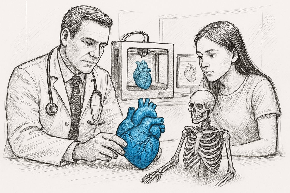
Imagine an operating room in 2026, where doctors prepare with a physical, patient-specific model in hand. 3D printed anatomical models have significantly changed both teaching and patient care.
This guide takes you from the technology behind to the latest trends, giving you everything you need to know. We cover processes, applications, materials and innovations.
Do you want to understand how the future of medicine, education and communication is being shaped? Read on and gain insight into best practices and the opportunities that await.
What is a 3D Printed Anatomical Model?
A 3D printed anatomical model is a physical representation of body parts, organs or pathological conditions created using advanced 3D printing technologies. These models are based on digital data, typically from CT or MRI scans, and are transformed via technologies such as FDM, SLA and SLS into detailed copies. Where traditional models are often standardized, 3D printed anatomical models enable unique individualization for each patient. Typical examples are skeletal parts, heart, liver or models of rare diseases. If you want a more thorough introduction to the concept, you can read more at What is an anatomical model? .
Definition and basic principles
A 3D printed anatomical model differs significantly from classic plastic models. Here, the starting point is real patient data, which opens up for precise, patient-specific solutions. The most commonly used technologies are FDM (Fused Deposition Modeling), SLA (Stereolithography) and SLS (Selective Laser Sintering). FDM is suitable for coarse structures such as bones, while SLA and SLS enable very fine details, ideal for organs and complex tissues. An example could be a 3D printed anatomical model of a heart with visible vessels, or a model of a tumor for training and planning.
Advantages compared to classic models
The advantages of a 3D printed anatomical model are many. First of all, it provides high precision and the ability to show even the smallest anatomical details. The model can be individually adapted to the patient's anatomy, which is not possible with standard models. This means better education, improved patient communication and more accurate surgical planning. According to recent studies, 73% of medical students experienced improved understanding when using a 3D printed anatomical model compared to traditional solutions.
| Advantage | Classic model | 3D printed anatomical model |
|---|---|---|
| Level of detail | Low | High |
| Individualization | No | Yes |
| Precision | Medium | High |
| Patient-specific use | No | Yes |
Development from 2020 to 2026
Since 2020, 3D printed anatomical models have evolved from being an experimental solution to becoming standard equipment in many hospitals and universities. New materials such as flexible polymers and biocompatible resins have made the models both more realistic and safe for clinical use. Technological breakthroughs have also significantly reduced production time. Today, 3D printed anatomical models are used not only in human medicine, but also in veterinary anatomy, where accurate animal models facilitate both teaching and treatment.
Examples of users and cases
The use of 3D printed anatomical models is rapidly spreading in the healthcare sector. Hospital departments use them for surgical planning, while universities and dental practices use them in teaching. A concrete example is a surgical team using a patient-specific model of the heart to plan a complex operation. Statistics show that the use of 3D printed anatomical models can reduce surgery time by up to 15%, according to current studies. This contributes to increased patient safety and better outcomes.
Limitations and challenges
Although 3D printed anatomical models have many advantages, they also present challenges. The cost of equipment and materials can be high, and production times vary from a few hours to several days depending on the complexity of the model. Not all materials are equally durable or realistic, and current technologies have limitations in resolution and finish. In addition, ethical and legal aspects must be taken into account, especially when patient data is used to produce 3D printed anatomical models. Quality assurance and documentation are therefore essential.
How to Make a 3D Printed Anatomical Model: Step-by-Step Guide
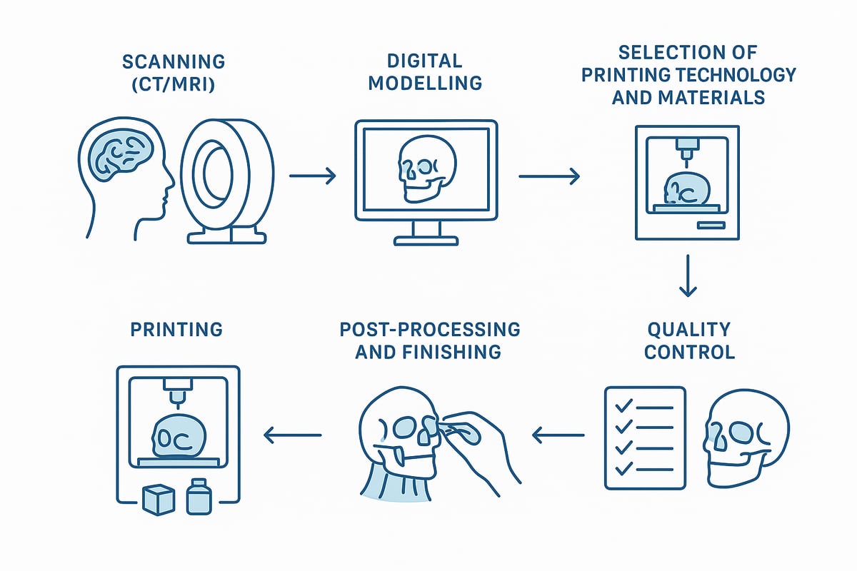
Producing a 3D printed anatomical model requires a systematic approach, where precision and quality are paramount. The process consists of six key steps that ensure that the model meets both professional and technical requirements. Here you will get a complete guide to each step, so you can create models that meet both educational and clinical needs.
1. Data collection and imaging
The first step in developing a 3D printed anatomical model is to collect accurate imaging data. Typically, CT or MRI scans are used, as these provide detailed images of the body's structures. Alternatively, 3D scanning can be used for surface models.
The choice of imaging modality depends on the desired model type. To achieve the necessary accuracy, the image resolution must be high and the slice distances small. It is important to ensure that all image data is handled in accordance with applicable data protection and patient confidentiality regulations.
- Use CT/MRI for internal structures
- 3D scanning for external parts
- Remember to obtain consent for patient data
Thorough imaging is the foundation for a successful 3D printed anatomical model.
2. Digital modeling and preparation
Once the image data is collected, digital modeling begins. Specialized software such as Mimics or 3D Slicer is used to segment and process the data so that it can be converted into a 3D printed anatomical model.
In this phase, the level of detail of the model is adjusted. Color codes and markers can be added to make the model more informative and user-friendly. It is important to export the files in the correct formats, typically STL or OBJ, which are supported by most 3D printers.
- Segment relevant anatomical structures
- Add necessary markings and textures
- Export in correct format
A well-executed digital preparation ensures that your 3D printed anatomical model will be both accurate and functional.
3. Choice of printing technology and materials
The choice of printing technology and materials has a great impact on the result when you make a 3D printed anatomical model. The most commonly used technologies are FDM, SLA and SLS, each of which has advantages and disadvantages.
- FDM is cheap and fast, but less detailed
- SLA provides high resolution, ideal for small structures
- SLS is suitable for complex and durable models
Materials such as PLA, ABS, resin and flexible polymers are chosen based on the purpose of the model. Durability and realism play a key role. For a deeper review of materials and technologies, you can read this guide to materials and technologies .
A well-thought-out combination of technology and material ensures that your 3D printed anatomical model meets both practical and legal requirements.
4. The 3D printing process itself
Once the model's digital file and materials are ready, the actual printing process begins. A 3D printed anatomical model is built layer by layer, and it is important to prepare the printer correctly.
Layer thickness and support structures must be adapted to the complexity of the model. During printing, the process is closely monitored to avoid errors such as warping or missing details. Printing time can vary from a few hours to several days, depending on the size and level of detail of the model.
- Prepare printer and materials
- Adjust layer thickness for optimal quality
- Monitor the process continuously
Effective control of the printing process is crucial for a successful 3D printed anatomical model.
5. Post-treatment and finish
After the print is complete, your 3D printed anatomical model must undergo post-processing. First, any supports are removed. The surface is polished, and the model can be painted or assembled if it consists of several parts.
Sterilization may be necessary, especially for clinical use. Some models have embedded chips or sensors to enable advanced functions. Proper post-processing extends the life of the model and increases its usefulness.
- Remove supports and polish the surface
- Paint or assemble the model parts
- Sterilize when necessary
A thorough finish ensures that your 3D printed anatomical model is ready for use in teaching or clinic.
6. Quality control and validation
The final step is quality control and validation of your 3D printed anatomical model. The model is compared to the original image data to ensure accuracy. Specialized measurement tools and feedback from experts are often used in this process.
Documentation of the process and possible CE marking is important, especially if the model is to be used in the healthcare sector. Systematic quality control minimizes errors and increases safety in use.
- Compare model with original data
- Get feedback from specialists
- Document and CE mark the model
Quality control is essential to ensure that your 3D printed anatomical model meets the highest standards.
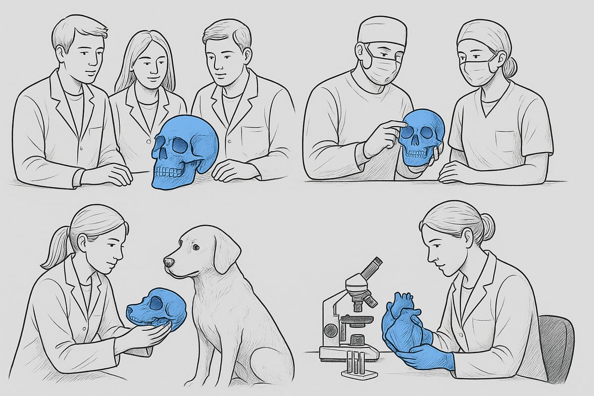
Areas of Application for 3D Printed Anatomical Models
3D printed anatomical models have quickly changed the way we work with anatomy in both the healthcare sector and educational environments. The possibilities range widely, from classical education to advanced surgical planning and research. Here we review the most important application areas where 3D printed anatomical models create value and innovation.
Medical education
In medical education, 3D printed anatomical models have revolutionized teaching. Students now have the opportunity to work with accurate, realistic models of organs, bones, and complex structures that were previously only available through cadavers.
The advantage of 3D printed anatomical models lies in the possibility of hands-on training without the ethical and logistical challenges that come with cadaver training. Statistics show that 60% more students prefer 3D printed models in teaching.
This development makes it easier to understand rare pathologies and variations, as the models can be adapted to specific needs. Read more about anatomical models for medical education , where you will find examples and inspiration for use in learning environments.
Surgical planning and simulation
For surgical teams, a 3D printed anatomical model can make a crucial difference in preparing for complex procedures. Patient-specific models, based on CT or MRI data, allow surgeons to practice procedures on an exact replica of the patient's anatomy.
This increases precision and reduces operating time because the surgeon has already experienced and visualized the challenges that may arise during the procedure. A concrete example is cardiac surgery, where a 3D printed anatomical model of the heart is used for simulation and planning. Statistics indicate up to 15% shorter operating time when using these models.
By integrating 3D printed anatomical models into clinical practice, better results and greater safety for the patient are achieved.
Patient communication and information
A 3D printed anatomical model has proven to be an effective tool in the dialogue between healthcare professionals and patients. When explaining diagnoses and treatment options, a physical model of the patient's own anatomy can provide a much more intuitive understanding.
Patients experience greater insight into their situation, which leads to increased compliance and better collaboration on the treatment process. Statistics show that 80% of patients feel better informed when 3D printed anatomical models are used during consultations.
The models can also be used to illustrate potential risks or benefits of different treatments, which strengthens trust between patient and practitioner.
Research and development
In research and development, 3D printed anatomical models open up testing and validation of new medical implants and devices. Researchers can design models that represent rare diseases or anatomical variations, providing unique opportunities to test and improve products.
Pharmaceutical research also benefits from 3D printed anatomical models, as they enable precise simulations of drug delivery or surgical techniques. The development process becomes more efficient and the results can be more easily transferred to clinical practice.
The use of 3D printed anatomical models in this context supports innovation and accelerates the introduction of new solutions to the market.
Veterinary medicine and other industries
Veterinary medicine has also embraced 3D printed anatomical models, both in education and treatment. Animal models are used to train students and plan surgeries, increasing the success rate of complex procedures.
Dental practices, physiotherapy and even artistic projects also use 3D printed anatomical models. One example is a complete horse skeleton, printed for use in veterinary education, where detail and customization make learning more effective.
This broad application emphasizes how versatile 3D printed anatomical models are and how the technology continues to spread to new fields of study.
Materials and Technologies: What Should You Choose?
The choice of materials and technologies is crucial for the quality, realism and functionality of a 3D printed anatomical model. Which material you choose depends on the purpose, and which technology is used can determine both the level of detail and durability.
Overview of the most commonly used materials
When choosing a material for a 3D printed anatomical model, you should consider strength, flexibility, color, and biocompatibility. PLA is popular for its environmental friendliness and ease of processing, while ABS offers more strength and durability. Resin provides high levels of detail and a smooth surface, which is especially useful for small structures. Nylon is often used for flexible or particularly robust models.
Here is a comparison table:
| Material | Strength | Flexibility | Biocompatible | Color selection |
|---|---|---|---|---|
| PLA | Medium | Low | Yes | Many |
| ABS | High | Medium | No | Get |
| Resin | Medium | Low | Yes* | Get |
| Nylon | High | High | Yes | Get |
*Please note that only medically approved resin is biocompatible.
You can see more examples of advanced materials at 3D dissection models and innovation .
Choice of printing technology
The choice of technology has a major impact on how a 3D printed anatomical model is experienced in practice. FDM (Fused Deposition Modeling) is the cheapest and fastest, but the level of detail is limited. SLA (Stereolithography) excels at high resolution and is suitable for fine details. SLS (Selective Laser Sintering) can create complex, durable models without the need for support structures.
According to recent statistics, 45% of the healthcare sector prefers SLA, especially when precision and small details are crucial. Consider technology based on purpose, economics and desired finish. It is important that the technology matches the requirements of your 3D printed anatomical model.
Special requirements for medical models
A 3D printed anatomical model for medical use must often meet special requirements for sterility and biocompatibility. The materials must be able to withstand sterilization without losing shape or properties. CE marking is a legal requirement for clinical use, and only certain materials are approved for patient contact.
Examples of approved materials include medical resin and sterilizable nylon. It is always recommended to document material selection and ensure that the entire manufacturing process complies with applicable legislation. This ensures that your 3D printed anatomical model is safe and reliable.
Environment, recyclability and materials of the future
Sustainability is gaining traction, also when it comes to 3D printed anatomical models. Several manufacturers now offer biodegradable or recyclable materials, such as PLA and certain types of nylon. Future innovations point towards the use of bioink and living cells, which could enable organ printing and even more realistic models.
Developments are expected to accelerate towards 2030, where environmentally friendly solutions and advanced materials will set new standards. Choosing sustainable materials supports both function and responsibility in the production of 3D printed anatomical models.
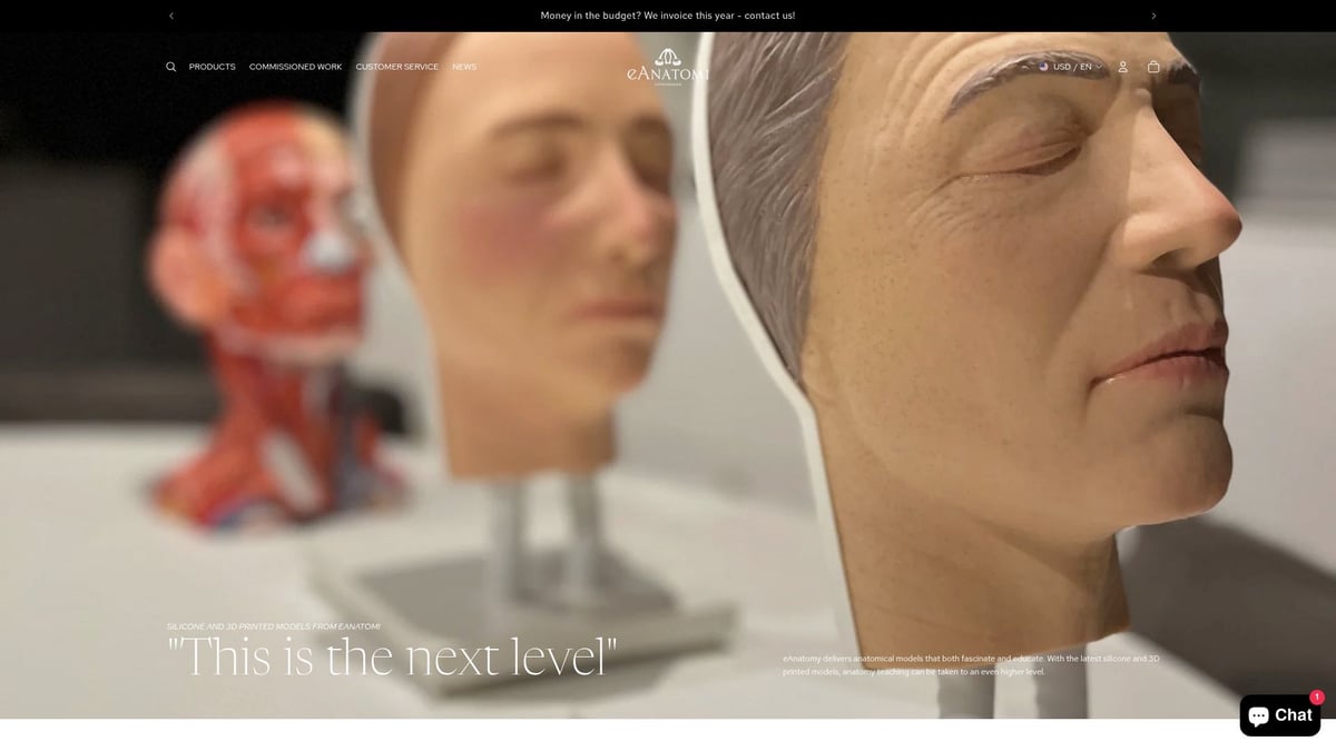
eAnatomy: Expert in Anatomical Models and 3D Printing
eAnatomi ApS is known as the leading supplier of anatomical models and educational materials in Denmark and the Nordic region. The company specializes in both classic solutions and modern 3D printed anatomical models, making them an obvious choice for the healthcare sector, universities and research institutions.
The company works closely with healthcare professionals to develop accurate and innovative models. This collaboration ensures that each 3D printed anatomical model meets clinical requirements and ethical standards. At the same time, eAnatomi offers the possibility of tailor-made solutions, so that the models can be adapted to specific needs within both medical education and patient communication.
The products range from detailed skeletal models and realistic organ models to specially designed pathological models and veterinary solutions. Many of the models are formalin-free, CE marked and made from advanced materials that ensure long durability and high realism. For example, 3D printed anatomical models are often used for surgical planning, where precision is crucial – read more about how 3D printed anatomical models improve surgical planning .
eAnatomi not only delivers to Danish institutions, but also to customers worldwide. With over 1000 institutions in Denmark and the Nordic region already using their products, the company is helping to set new standards for the use of 3D printed anatomical models in both teaching, research and clinical practice.
Future Trends and Innovations in 3D Printed Anatomy
The development of 3D printed anatomical models is moving rapidly, and 2026 is shaping up to be a groundbreaking year. New technologies and methods are significantly changing how the healthcare sector works with anatomy, education and patient care. Here you will get an overview of the most important trends and innovations that are shaping the future of 3D printed anatomical models.
Artificial intelligence and automated modeling
Artificial intelligence is playing an increasingly important role in the development of 3D printed anatomical models. AI algorithms can now segment and process image data faster than ever before. This means that models can be designed with far less manual effort, minimizing the risk of error.
New software solutions integrate AI directly into the workflow. This makes it possible to generate accurate, patient-specific models with just a few clicks. According to industry data, AI can shorten modeling time by up to 60 percent. For both hospitals and educational institutions, this means faster access to accurate 3D printed anatomical model solutions.
3D printing with living tissue and bioink
One of the most revolutionary innovations is 3D printing with living tissue, also known as bioprinting. This uses bioink containing living cells to create structures such as organs or tissues that can potentially be used for transplantation or research.
Although the technology is still under development, the first clinical applications are expected within a few years. Researchers are working intensively to solve challenges with tissue maturation and integration in the body. The development of 3D printed anatomical models with bioink opens up completely new possibilities for the treatment of complex diseases and personalized medicine.
Integration with VR and AR
Virtual and augmented reality (VR and AR) are increasingly being integrated with physical models. When a 3D printed anatomical model is combined with VR, healthcare professionals can train in realistic, interactive environments, improving understanding of complex anatomical structures.
For example, VR is currently used for surgical training, where the doctor both sees and feels the model during simulation. This combination significantly increases learning outcomes and reduces errors in practice. AR technology makes it possible to layer digital layers on top of physical models, providing even deeper insight.
On-demand production and decentralization
An important trend is decentralization, where hospitals and clinics establish their own 3D labs for on-demand production. This shortens the delivery time of a 3D printed anatomical model and allows for immediate adaptation to the patient's needs.
A good example is the 3D Lab at Aarhus University Hospital , where specialists produce models for surgical planning and teaching directly on site. However, this development requires quality control and standardization so that the models meet clinical requirements.
Future education and patient care
In the coming years, 3D printed anatomical models are expected to become standard in both education and patient treatment. The market is growing by 20 percent annually, and new solutions focus on personalization and digitalization.
Students and healthcare professionals will have access to more realistic, patient-specific models that can be combined with digital tools. The future points to a tight integration of technologies, with 3D printing trends for 2026 showing how innovation continues to transform the possibilities for both learning and treatment.
Once you have an overview of the technology, possibilities and future trends for 3D printed anatomical models, you may have questions about how to best choose materials, technology and supplier. At eAnatomi, we have specialized in developing innovative, CE-marked solutions that make it easy for you to integrate 3D printed anatomy into both teaching and clinical practice. If you want to delve deeper into the subject, see concrete examples or find the right model for your needs, you can Read more here
0 comments