Skip to product information
Pædagogisk hjerne, 2 dele - eAnatomi
1/3

Educational brain, 2 parts

Varenr.: A612
$123.00
Shipping calculated at checkout.
In stock

Pickup available at Frederiksberg

Usually ready in 4 hours

Educational brain, 2 parts

Default Title

Frederiksberg

Pickup available, usually ready in 4 hours

Helgesvej 12
2000 Frederiksberg
Denmark

+4529919911
Beskrivelse

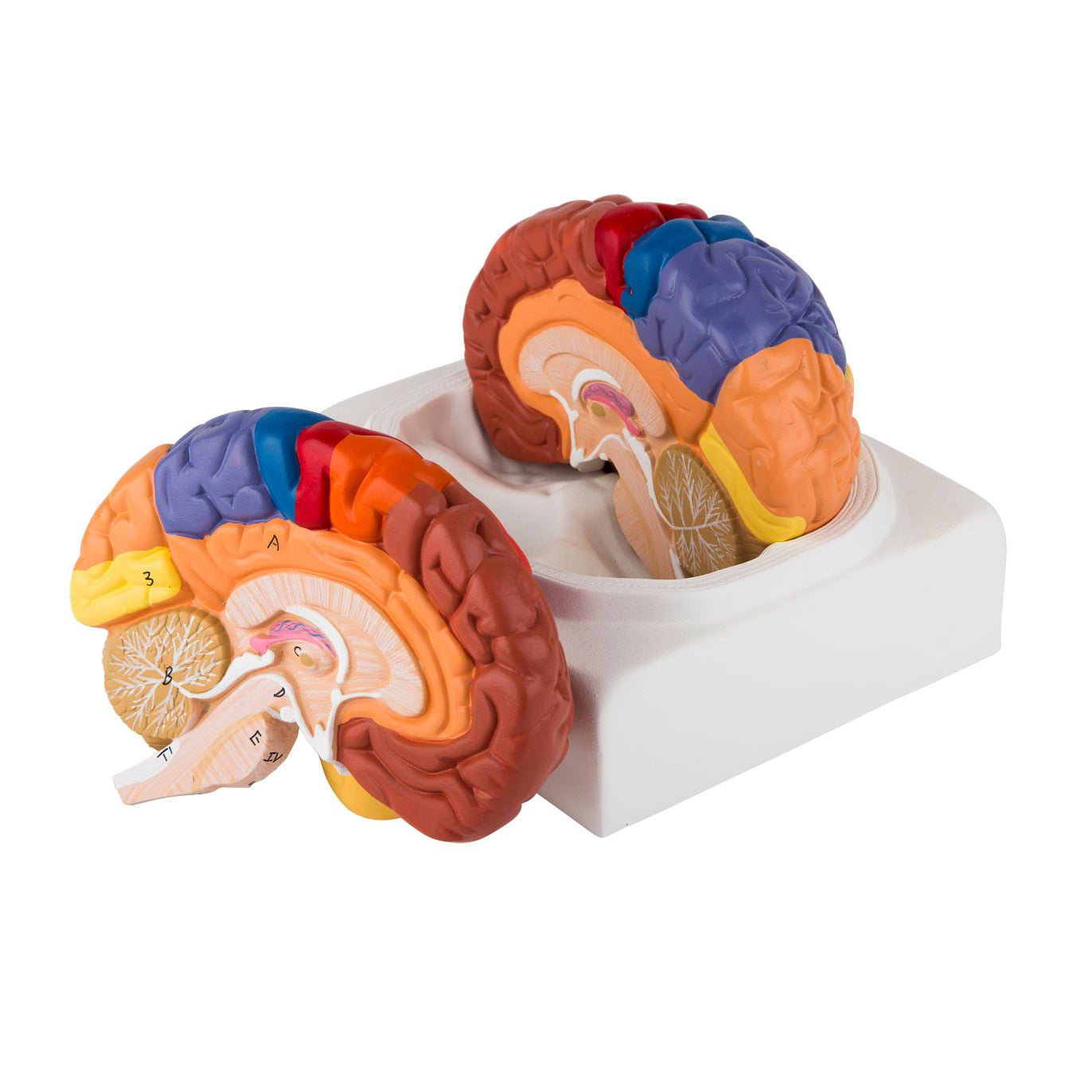
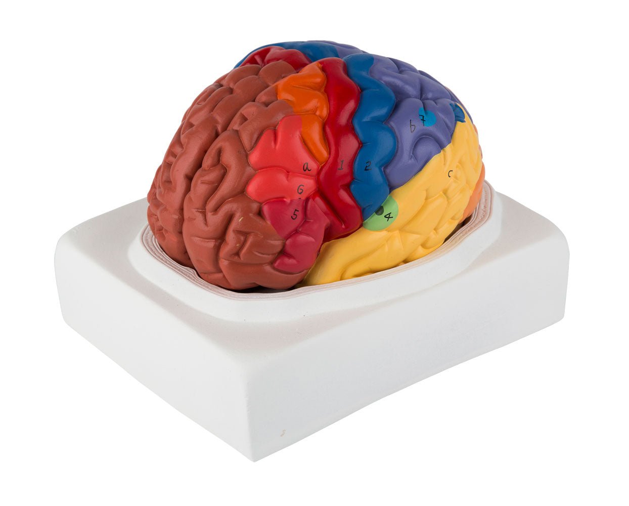
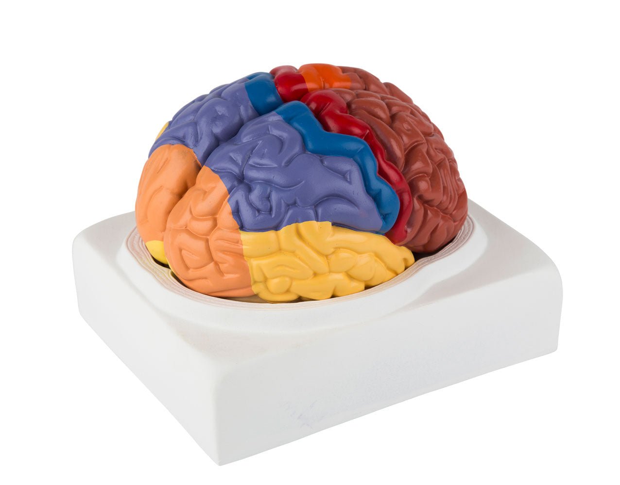
If you are looking for a simple brain model which shows the most important areas of the brain in educational colours, this model is ideal. It is an inexpensive alternative to our brain models with educational colors in higher price ranges.


The model is cast in very hard and robust plastic. Unlike many of our other brain models, which are molded in hollow and flexible plastic, this model's material cannot be pinched or moved a bit. Some would argue that this makes it less pleasant to touch and work with when it needs to be taken apart and studied. Others will think that it has no meaning.


The model is split in the middle, and the 2 parts are not held together via metal pins or magnets. The size of the model corresponds to the brain of an adult person. Below you can read more about the anatomical details such as the colored areas and the limbic system. The model is supplied on a stand in white plastic (the area of ​​the stand where the brain model lies is shaped like the inner skull base, which can be seen in one of the pictures on the left).


Important anatomical structures and colored areas are numbered on the model. With the purchase, an overview with naming is included, cf. the mentioned numberings. The naming is only prepared in English.

NB: The numbering and naming are indicative. Therefore, be critical in your use, as figures for an anatomical structure may, for example, be located on the border of another structure.

Anatomisk set

Anatomically, the model shows the human brain, which can generally be divided into the cerebrum (cerebrum), the cerebellum (cerebellum) and the brain stem (truncus encephali).


These 3 structures are clearly separated via different colors, but the difference between gray and white matter cannot be seen on this model (however seen in the cerebellum).


In the cerebrum (telencephalon and diencephalon), the lobes of the brain, as well as the thalamus and hypothalamus (and the pituitary gland) are primarily seen


In the cerebellum, the vermis cerebelli and the cerebellar hemispheres (hemisperium cerebelli) are seen


The brainstem shows its 3 parts (the midbrain, the pons and the medulla oblongata) as well as the apparent origins of the cranial nerves using Roman numerals (also called the cranial nerves)


Other structures are also seen, such as the brainstem, the fornix, a bit of the ventricular system and the first 2 cranial nerves (the olfactory and optic nerves), which do not originate from the brainstem


Colored areas


The cerebrum, cerebellum and brainstem are seen in different colours. In addition, important areas in the cerebrum are shown with different colors. These are, for example, the different lobes of the brain, the primary motor area in the frontal lobe, the area for the primary sensory function in the parietal lobe and the area for primary visual perception in the occipital lobe.


The limbic system


Many of our customers ask about the limbic system in connection with the purchase of brain models. Hence this description.


The limbic system includes various anatomical structures in the central nervous system (CNS), and is primarily responsible for emotional functions such as anxiety, aggressiveness, mood, memory and social adaptability. Clinically, it is therefore often related to psychiatric disorders.


The limbic system includes, among other things amygdala, hippocampus, gyrus parahippocampalis, hypothalamus, fornix, corpus mammillare, the prefrontal cerebral cortex and the monoaminergic systems of the brainstem. The list is quite a bit longer - especially because numerous fiber connections connect the limbic structures. Many customers ask in particular about the amygdala and hippocampus (which is why they are mentioned first in this section).


NB: In this brain model, neither the amygdala nor the hippocampus can be seen, but some of the other limbic structures can be seen (e.g. the fornix).


The amygdala is involved in anxiety and emotional coloring of sensory impressions. It lies as an almond-shaped nucleus IN FRONT of the hippocampus in the anterior pole of the temporal lobe (amygdala and hippocampus are therefore separate).


The hippocampus is involved in memory. It lies as an irregular twisted structure in the medial part of the temporal lobe.

If one of these 2 structures is to be seen clearly on a brain model, the brain tissue must be shown in a so-called frontal/coronal section through the temporal lobe. The frontal incision roughly corresponds to the incision direction "from ear to ear".


Because the amygdala is IN FRONT of the hippocampus (roughly speaking further forward "toward the forehead"), both of these structures can only be seen on a brain model if the model includes at least 2 frontal sections through the temporal lobe - or if the brain model is partially transparent.


We have not yet seen a brain model that shows 2 cuts through the temporal lobe, so that both the amygdala and the hippocampus are seen. In our range, on the other hand, we have a partially see-through brain model in the highest price range, which shows both structures.


All brain models in our range can be separated into different parts. All models (both with and without educational colors) that can be separated into 4 or more parts show the hippocampus. On almost all of these models, the hippocampus is also numbered and named on an overview that can be downloaded from the product descriptions of the brain models.

Bevægelsesmæssigt
Klinisk set

Clinically, the model is ideal for understanding the problems (symptoms) and damage caused by lesions in the colored areas of the cerebrum on the model. This can, for example, concern disturbances in movements, sense of touch, language and vision.


The model can also be used to understand other lesions or disorders in the areas of the brain that are seen on the model.

KUNDESERVICE

bestillingsvarer

Er produktet angivest som bestillingsvare betyder det at produktet er i en størrelsesorden eller et så højt kvalitetsniveau, at produktet kun fremskaffes på bestilling. Leveringstiden kan variere, men prisen vil altid forblive den samme! Kontakt os for mere information, hvis du ønsker at bestille.

Priser og betalingsmetoder
  • Priser er angivet i DKK inkl. moms.
  • Du kan betale med;
    - EAN nr.
    - MobilePay
    - Visa, Mastercard
Fortrydelsesret & returnering
  • Fortrydelsesret er 14 dage fra levering.
  • Returporto er 60,- ved køb af label hos os (sendes som PDF fil via email) eller 0,- ved personlig returnering på vores adresse.
  • Se vores handelsbetingelser her
Levering / afhentning

Er varen angivet som på lager, befinder den sig fysisk på vores adresse på Frederiksberg og kan derfor afsendes eller afhentes. Vælg ved bestilling hvad du ønsker.

Ønsker du at afhente varen, kan det ofte finde sted umiddelbart efter bestilling, men skal aftales med os. Ønsker du afsendelse sker dette via postnord og ofte samme dag som bestillingen modtages.

Er varen angivet som 'sendes direkte fra fabrikken', er leveringstiden oftest 2-5 hverdage, men afhænger af fabrikkens lagerstatus. Ønsker du mere præcis information kan du altid kontakte os før bestilling.

Fakta om eAnatomi
  • Stiftet i 2004 som enkeltsmandsforretning og omdannet til ApS i 2019
  • 100% ejet af Christian Birksø som også står for den daglige drift.
  • Udvikler og markedsfører både originale produkter, designet og produceret af eAnatomi, samt forhandler mange internationale varemærker.
  • Sælger til både private og erhverv i Danmark og udland.

Andre produkter udvalgt specielt til dig

Vores anatomiske varesortiment

Lodtrækning om gratis produkter

Alle modtagere af vores nyhedsbrev er med i lodtrækninger om gratis produkter. Skriv dig op i dag og vær med!