Skip to product information
Naturtro hjerne, 15 dele - eAnatomi
1/5

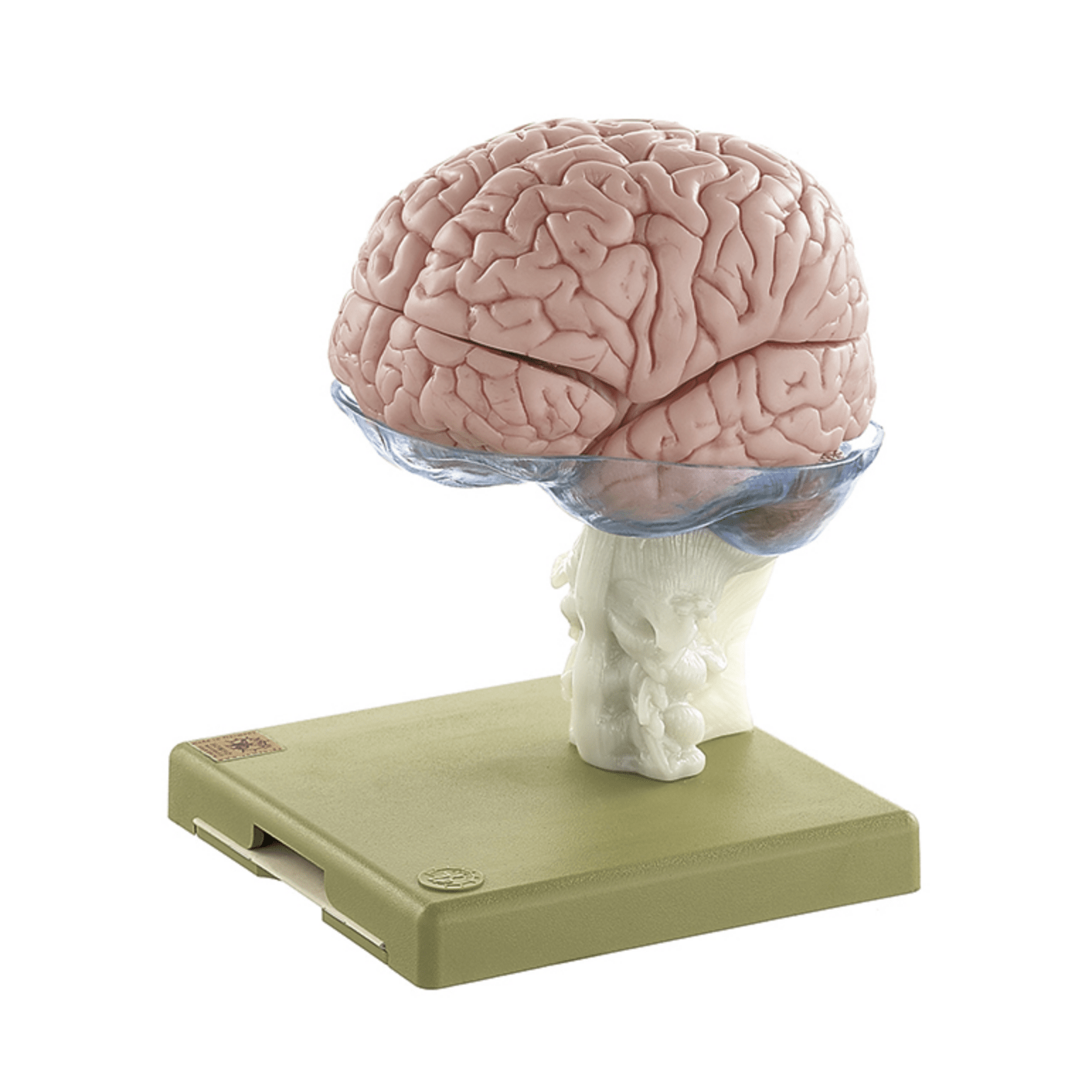
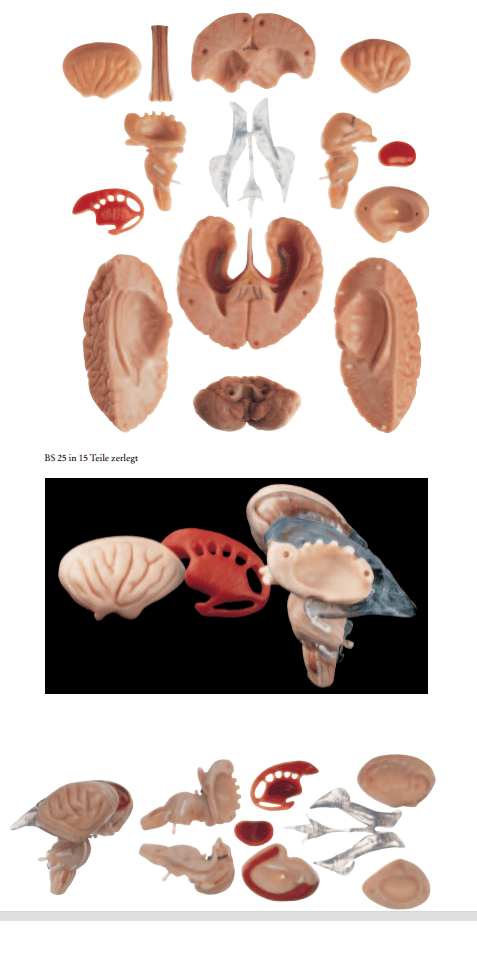
Brain, lifelike and advanced anatomical model in 15 parts

6.605,00 kr
Taxes included. Shipping calculated at checkout.
Out of stock
Description

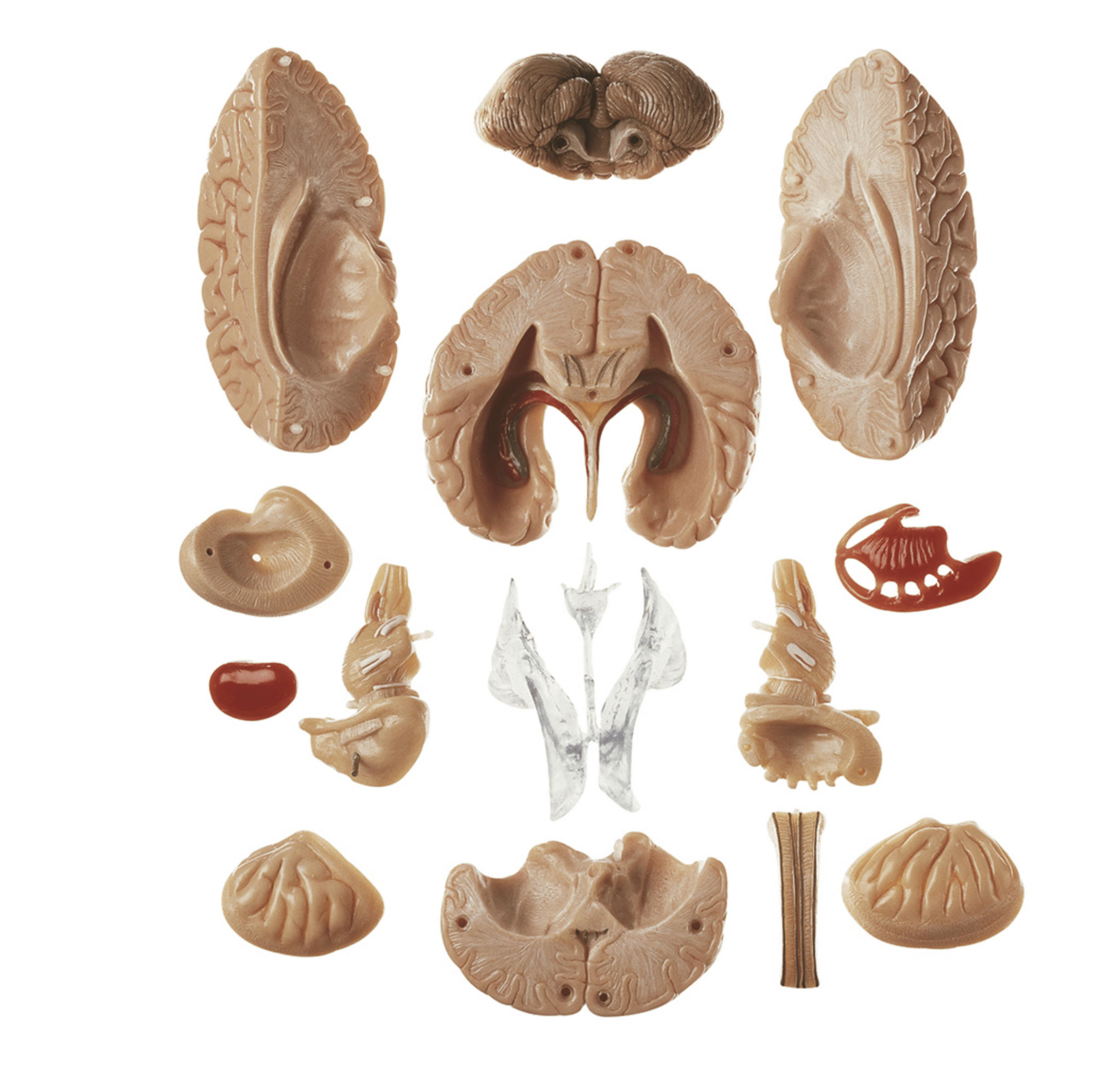
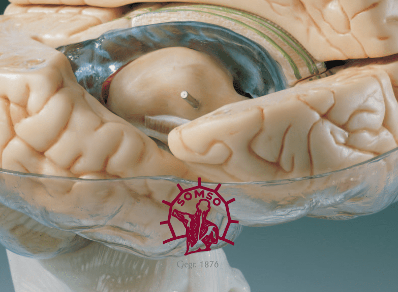
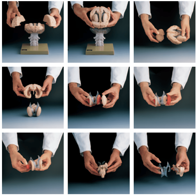
This brain model offers highly accurate reproductions of the brain's anatomical details, colored internal structures and can be separated into 15 parts.


The brain model was developed with the help of Prof. Dr. JW Rohen, Department of Anatomy at the University of Erlangen. It is produced in SOMSO plastic by the manufacturer SOMSO, which is world-renowned for very high quality. This means quality materials, a sense of accuracy and longevity.

In other words: World-class craftsmanship.


The mentioned SOMSO plastic makes the material flexible because it can be moved slightly. This makes the model pleasant to touch and work with when it needs to be taken apart and studied.


The model's 15 parts are held together via nylon pins. Below you can read more about the anatomical details such as the colored areas and the limbic system. The size of the model corresponds to the brain of an adult person. Incl. the cervical spine is the dimensions are 23 x 15 x 18 cm,

and the weight is 1.8 kg. It is delivered on a special stand, which consists of 3 parts: A transparent plate on which the model rests as well as the cervical spine and a green plate at the bottom.


The model is not numbered, but an overview has been made in which it is described what you can see in general on the model.

Anatomically speaking

Anatomically, the model shows the human brain, which can generally be divided into the cerebrum (cerebrum), the cerebellum (cerebellum) and the brain stem (truncus encephali).


These 3 structures are clearly separated via different color tones, and the difference between gray and white matter can be clearly seen on this model.


In the cerebrum (telencephalon and diencephalon), the lobes of the brain, as well as the thalamus and hypothalamus (and the pituitary gland) are primarily seen


In the cerebellum, the vermis cerebelli and the cerebellar hemispheres (hemisperium cerebelli) are seen


In the brainstem you can see its 3 parts (the midbrain, the pons and the medulla oblongata) as well as the apparent origin of the cranial nerves (also called the cranial nerves)


Other structures such as the brain stem, fornix, ventricular system and the first 2 cranial nerves (the olfactory and optic nerves) are also seen, which do not originate from the brainstem


The pictures on the left show how the model can be separated into 15 parts. This makes it possible to study the internal structures of the brain, and many of these are seen in 3 dimensions. These are, for example, the corpus callosum, corpus striatum, insula, nucleus lentiformis and the brainstem.


As previously mentioned, the cervical spine is seen as part of the stand. You can see the bone tissue and ligaments, but it is not suitable for studying the anatomical details of the cervical spine.


Colored areas


On this model, only some selected internal structures are colored (see images on the left). For example, the ventricular system appears transparent with a faint bluish tinge (the ventricular system can also be taken out of the model completely). The colors make it easy to identify important anatomical areas.


The limbic system


Many of our customers ask about the limbic system in connection with the purchase of brain models. Hence this description.


The limbic system includes various anatomical structures in the central nervous system (CNS), and is primarily responsible for emotional functions such as anxiety, aggressiveness, mood, memory and social adaptability. Clinically, it is therefore often related to psychiatric disorders.


The limbic system includes, among other things amygdala, hippocampus, gyrus parahippocampalis, hypothalamus, fornix, corpus mammillare, the prefrontal cerebral cortex and the monoaminergic systems of the brainstem. The list is quite a bit longer - especially because numerous fiber connections connect the limbic structures. Many customers ask in particular about the amygdala and hippocampus (which is why they are mentioned first in this section).


NB: In this brain model, the hippocampus can be seen as well as some of the other limbic structures such as the fornix - but not the amygdala.


The amygdala is involved in anxiety and emotional coloring of sensory impressions. It lies as an almond-shaped nucleus IN FRONT of the hippocampus in the anterior pole of the temporal lobe (amygdala and hippocampus are therefore separate).


The hippocampus is involved in memory. It lies as an irregular twisted structure in the medial part of the temporal lobe.

If one of these 2 structures is to be seen clearly on a brain model, the brain tissue must be shown in a so-called frontal/coronal section through the temporal lobe. The frontal incision roughly corresponds to the incision direction "from ear to ear".


Because the amygdala lies IN FRONT of the hippocampus (roughly speaking further "toward the forehead"), both of these structures can only be seen on a brain model if the model includes at least 2 frontal sections through the temporal lobe - or if the brain model is partially transparent.


We have not yet seen a brain model that shows 2 cuts through the temporal lobe, so that both the amygdala and the hippocampus are seen. In our range, on the other hand, we have a partially see-through brain model in the highest price range, which shows both structures.


All brain models in our range can be separated into different parts. All models (both with and without educational colors) that can be separated into 4 or more parts show the hippocampus. On almost all of these models, the hippocampus is also numbered and named on an overview that can be downloaded from the product descriptions of the brain models.

Movement-wise
Clinically speaking

Clinically, the model is ideal for understanding lesions and disorders in specific internal structures. This is, for example, hydrocephalus, because the entire ventricular system can be taken out of the model and studied in detail. Another example is Parkinson's disease, which involves pathological changes in the striatum.


The model can also be used to understand many other lesions such as epilepsy, brain tumors, lesions involving cranial nerves and sclerosis (multiple sclerosis).


Although the brain's blood supply is not visible on the model, it can also be used to understand apoplexy (stroke).

CUSTOMER SERVICE

custom-made items

If the product is stated as a made-to-order item, it means that the product is of such a size or such a high quality level that the product is only available on order. Delivery time may vary, but the price will always remain the same! Contact us for more information if you wish to order.

Prices and payment methods
  • Prices are stated in DKK including VAT.
  • You can pay with;
    - EAN no.
    - MobilePay
    - Visa, Mastercard
Right of withdrawal & return
  • The right of withdrawal is 14 days from delivery.
  • Return postage is 60,- when purchasing a label from us (sent as a PDF file via email) or 0,- when returning in person to our address.
  • See our terms and conditions here
Delivery / pickup

If the item is listed as in stock , it is physically located at our address in Frederiksberg and can therefore be shipped or picked up. When ordering, choose what you want.

If you wish to pick up the item, this can often take place immediately after ordering, but must be agreed with us. If you wish to have it shipped , this will be done via Postnord and often the same day the order is received.

If the item is listed as ' shipped directly from the factory ', the delivery time is usually 2-5 business days, but depends on the factory's stock status. If you want more precise information, you can always contact us before ordering.

Facts about eAnatomy
  • Founded in 2004 as a sole proprietorship and converted to ApS in 2019
  • 100% owned by Christian Birksø who is also responsible for the daily operations.
  • Develops and markets both original products, designed and produced by eAnatomi, and distributes many international brands.
  • Sells to both private and commercial customers in Denmark and abroad.

Other products selected especially for you

Our anatomical product range

Raffle for free products

All recipients of our newsletter are entered into raffles for free products. Sign up today and be in the running!