Milt, bugspytkirtel, tyndtarm og nyrer, 3 dele - eAnatomi

Spleen, pancreas, small intestine and kidneys, anatomical model in 3 parts

€342,95
Sale price  €342,95 Regular price 
Skip to product information
Milt, bugspytkirtel, tyndtarm og nyrer, 3 dele - eAnatomi
1/7

Spleen, pancreas, small intestine and kidneys, anatomical model in 3 parts

SKU: 1000310

This product cannot be ordered online. Please contact us for more information.

Description

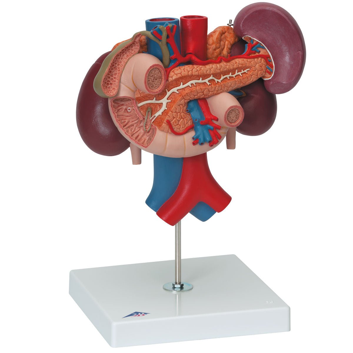
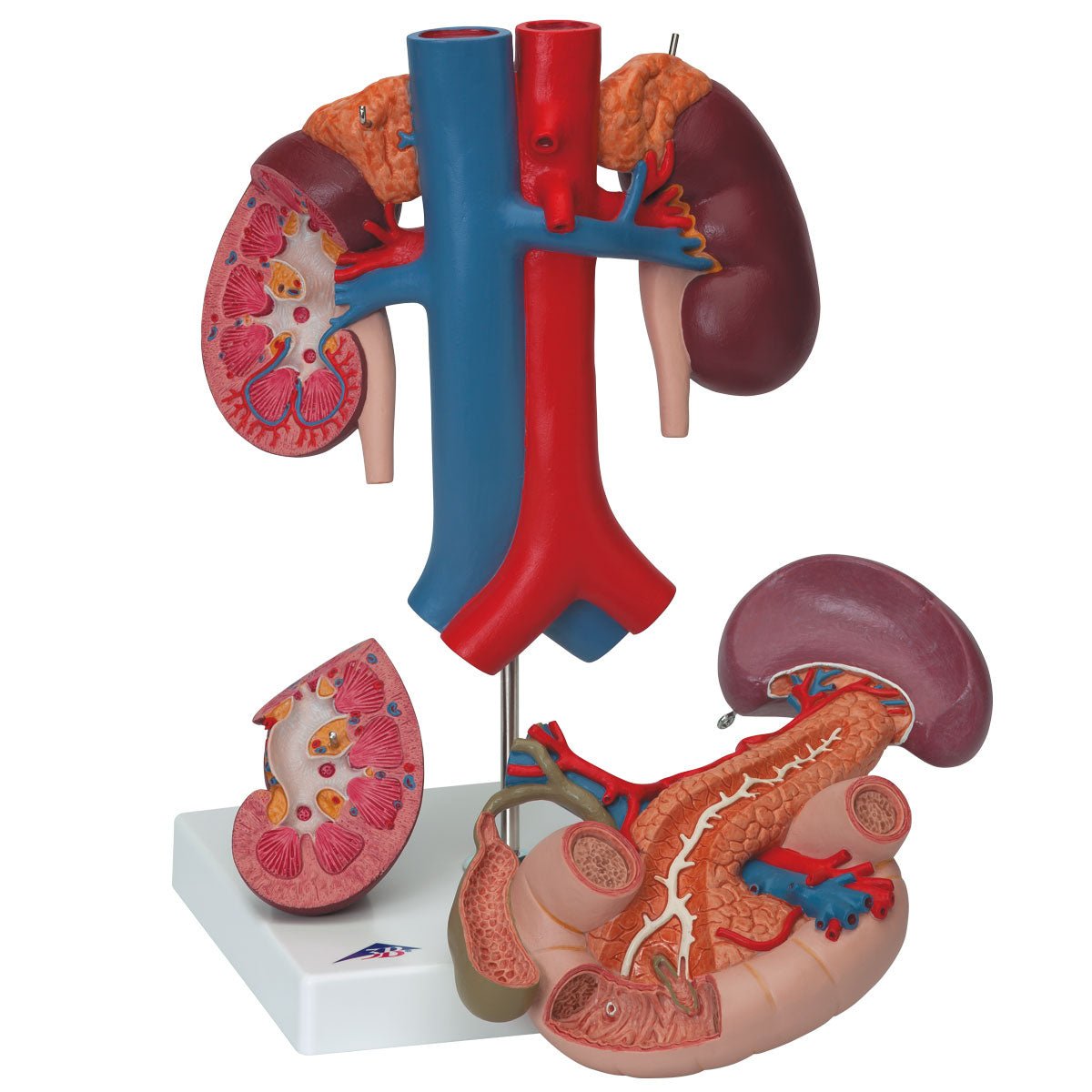
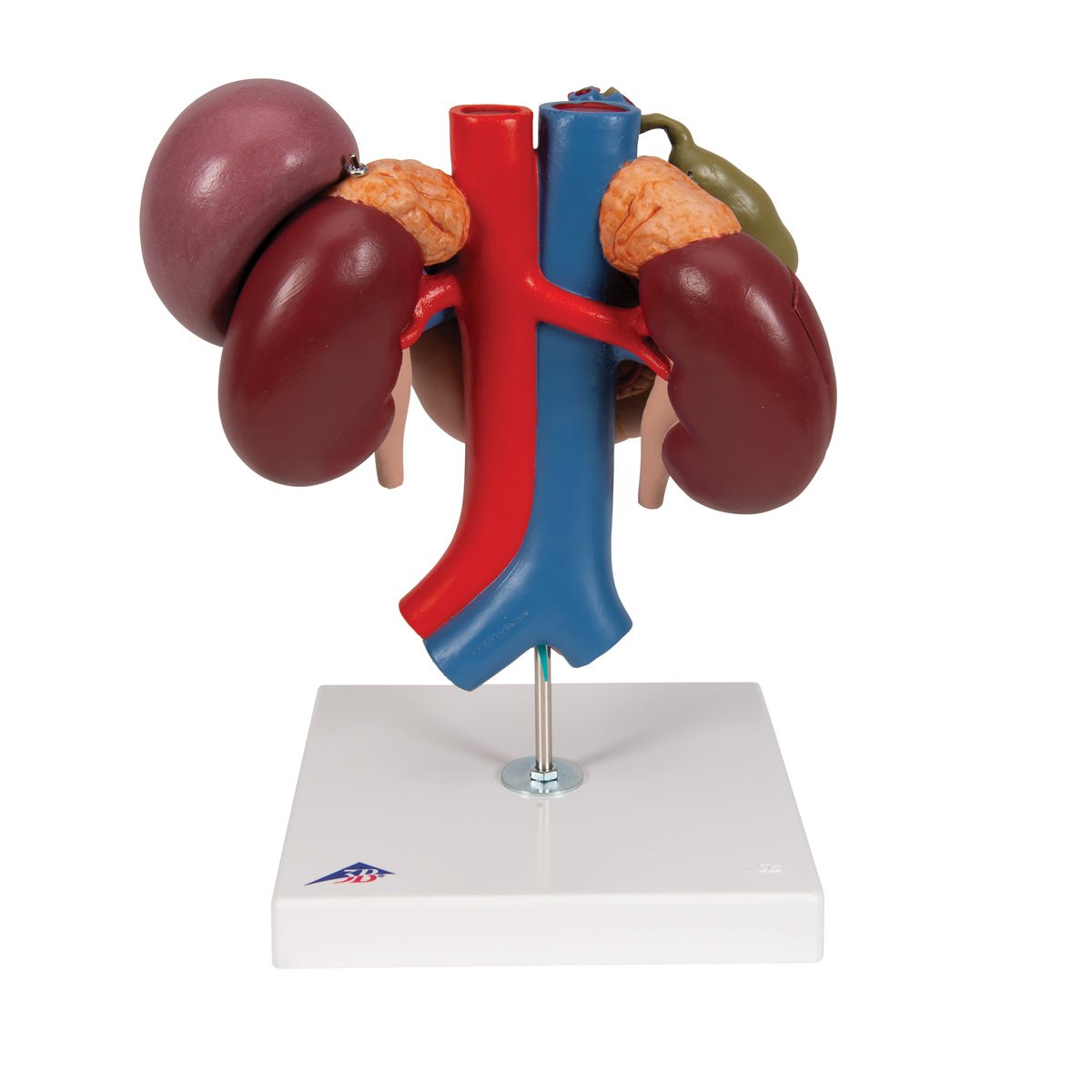
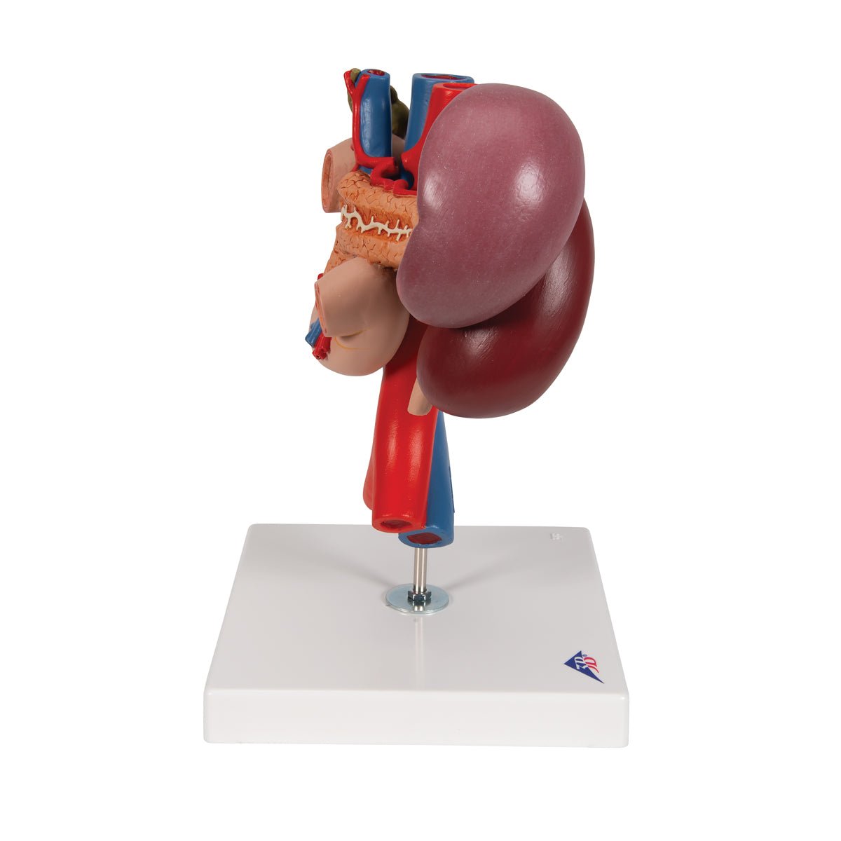
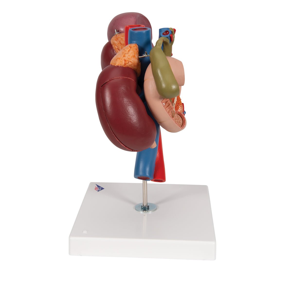
If you are looking for a model that gives a good overview of the relationships between the organs in the upper part of the abdominal region, and at the same time reproduces the individual organs in a fine degree of detail, we recommend this one.


This detailed model shows the relationship of the duodenum and pancreas to other organs such as the gallbladder, spleen and kidneys.


The model is produced in natural size, weighs 0.9 kg and measures 24 x 18 x 29 cm. It can be separated into 3 parts and is delivered on a white stand.

Anatomically speaking

Anatomically speaking, this model, with its spatial structure, provides a good understanding of how the organs are located in the abdominal cavity. The model shows the following organs:


Duodenum
The pancreas
Gallbladder and bile ducts
The spleen
Both kidneys incl. beginning of the ureters and adrenal glands
The largest arteries and veins in the region


Several of the organs are partially open, which gives an educational insight into important internal structures. You can see details such as:


Intestinal lumen of the duodenum incl. duodenal papilla major and minor
Pancreatic duct
The bile ducts (the extrahepatic bile duct system)
The interior of the gallbladder
The special tissue of the kidney with marrow, cortex, renal pelvis and blood vessels (only in the right kidney)


Of vessels, the model shows the largest arteries and veins passing through areas, including:


A. mesenteric superior and V. mesenteric superior
V. mesentica inferior
A. splenic and V. splenic
A. hepatica communis, A. hepatica propria and A. gastroduodenalis
Aorta
Inferior vena cava

Movement-wise
Clinically speaking

Clinically speaking, the model is ideal for understanding diseases/disorders such as stones in the bile ducts/gallbladder, inflammation of the gallbladder, pancreatitis and pancreatic cancer. It is also ideal for understanding diseases/disorders in the kidneys such as kidney stones, hydronephrosis and kidney trauma.


Furthermore, the model can be used to understand the purpose of imaging examinations in the region and treatments such as cholecystectomy.

Contact us

Our anatomical product range

Raffle for free products

All recipients of our newsletter are entered into raffles for free products. Sign up today and be in the running!