Microscopic anatomy

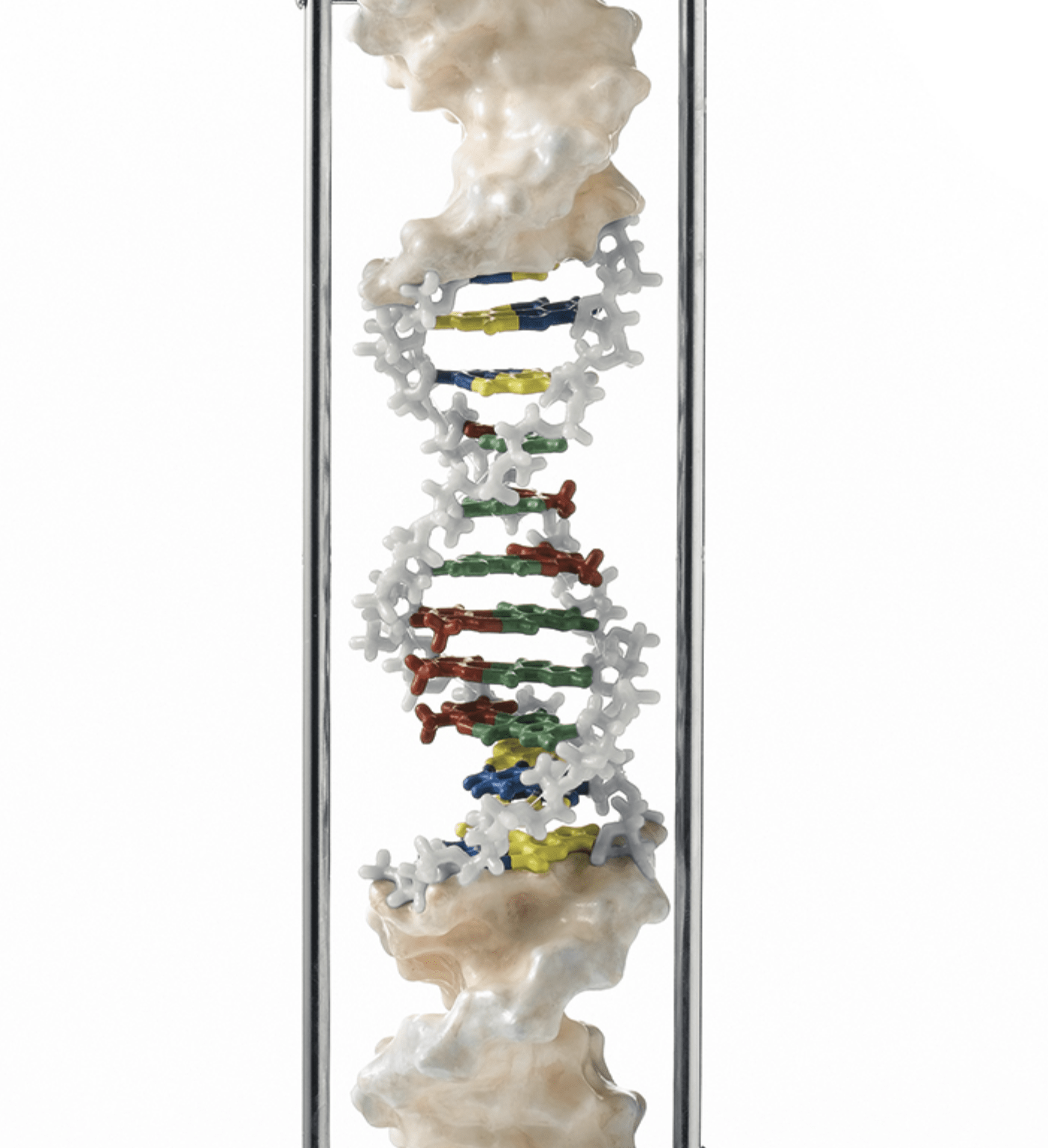
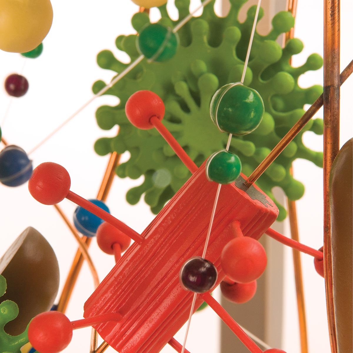
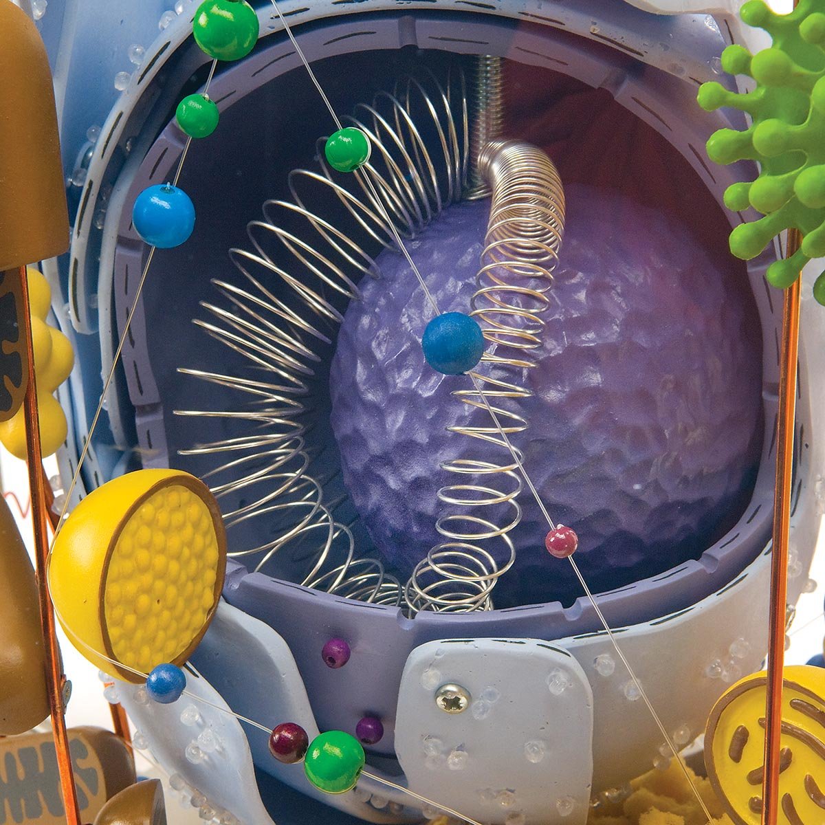
Human tissue is made up of cells, and the study of microscopic anatomy is essential for understanding both healthy and sick humans. Here we have collected our models that illustrate different types of tissue from healthy humans seen through an optical or electron microscope, as well as models of DNA; the human building block.

Skip to results list

Active filters:

18 items
Column grid
Column grid

Filter

Active filters:

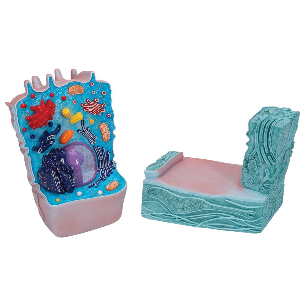
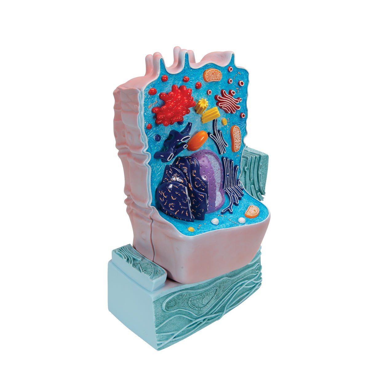
  • Cell, anatomical model

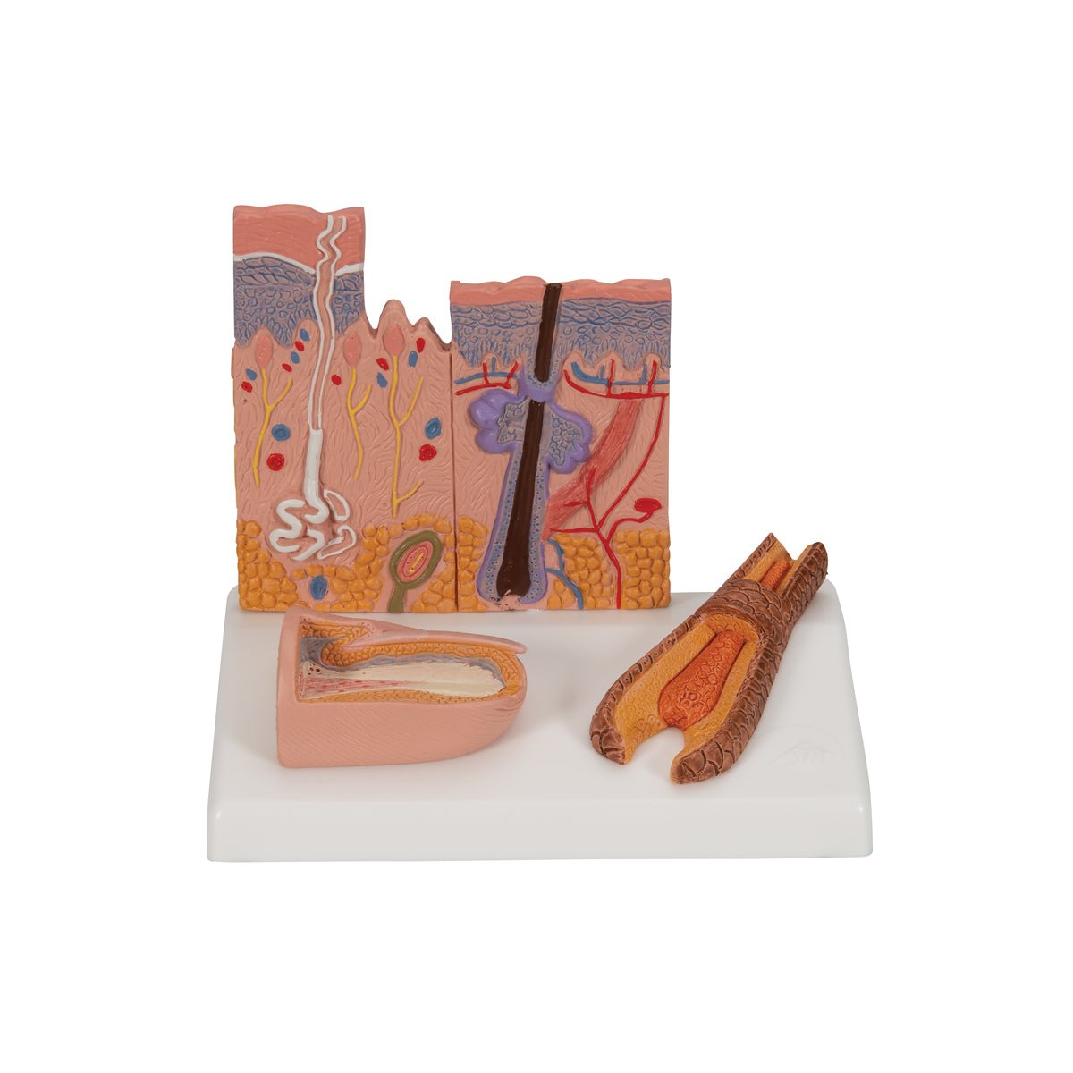
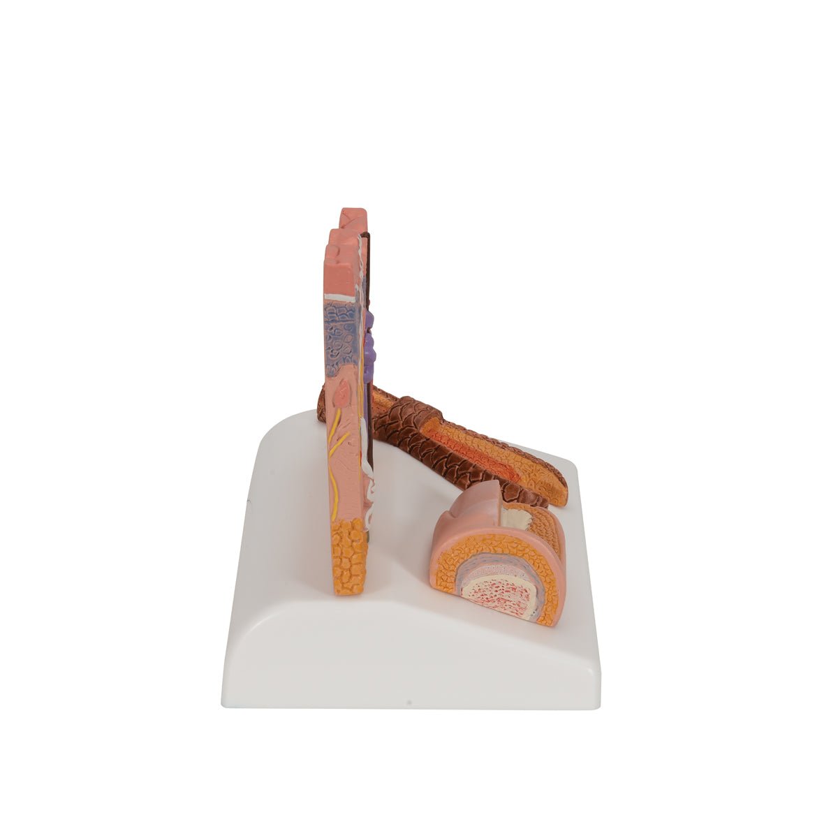
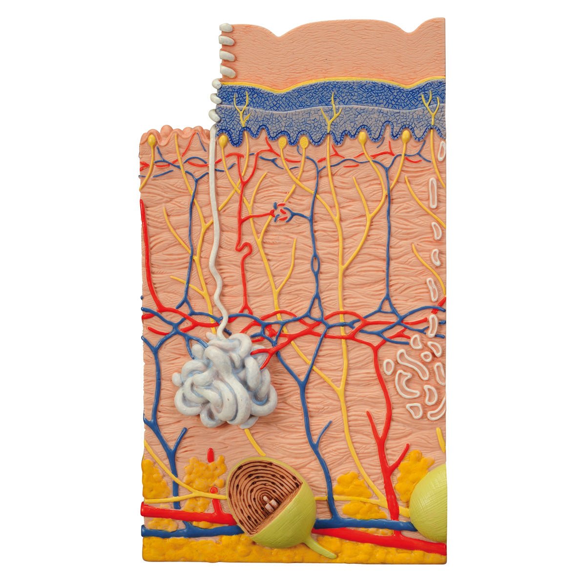
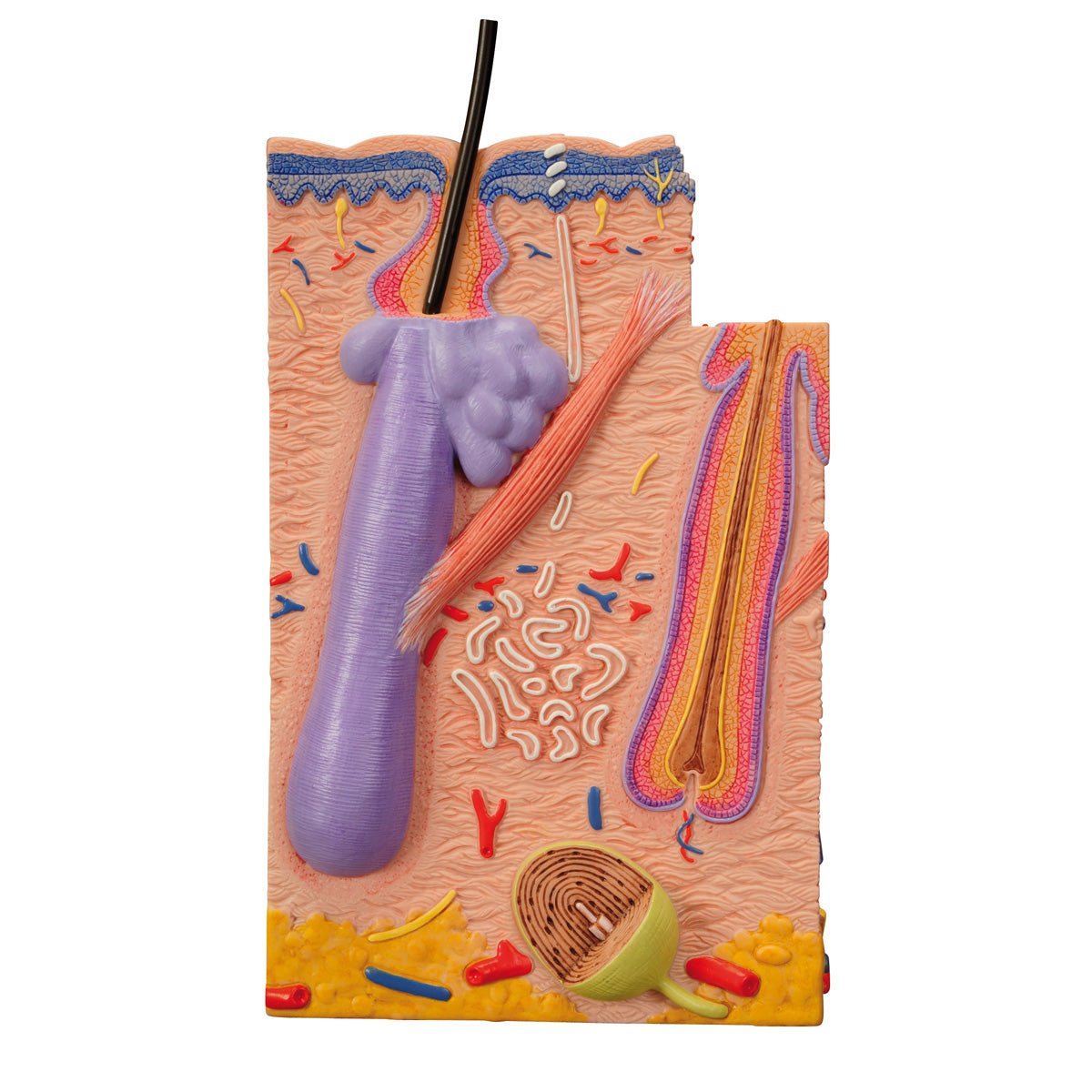
  • Skin, nail and hair root, enlarged anatomical model

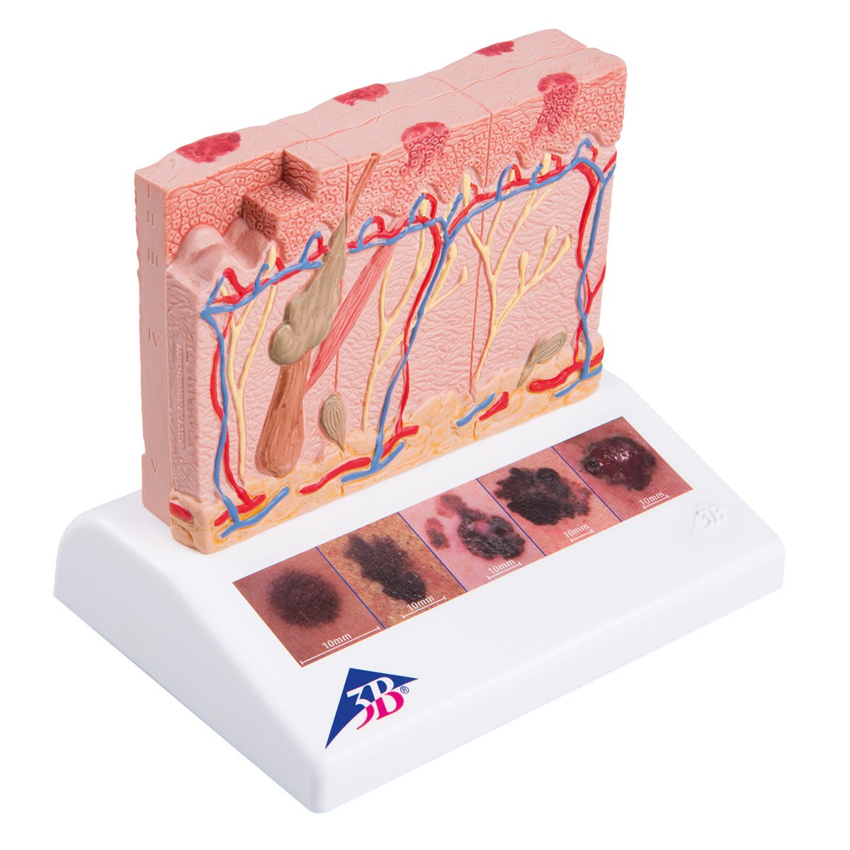
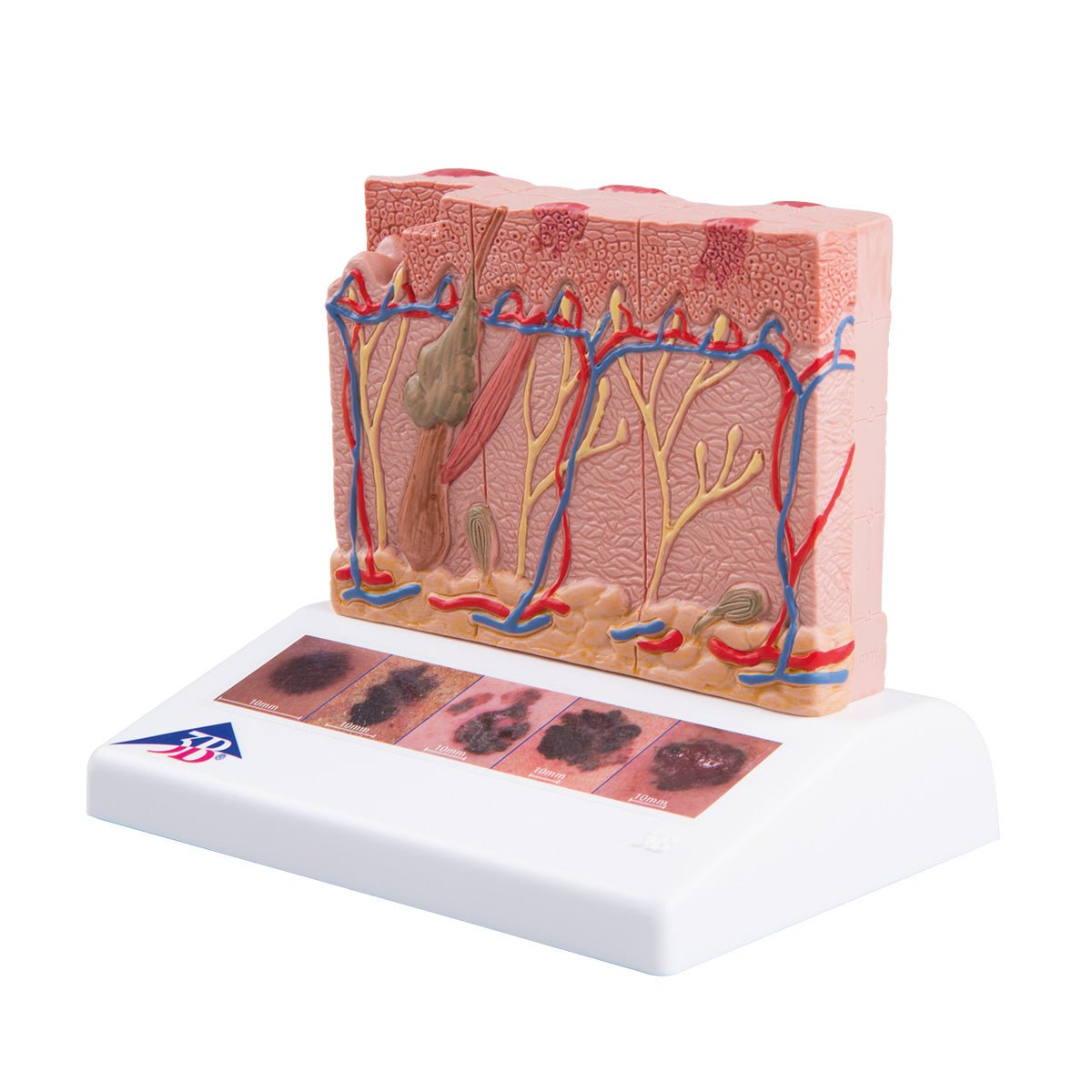
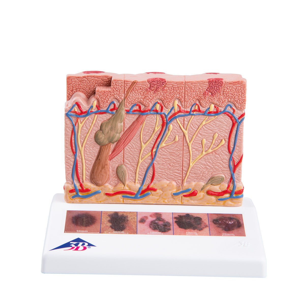
  • Melanoma, enlarged anatomical model

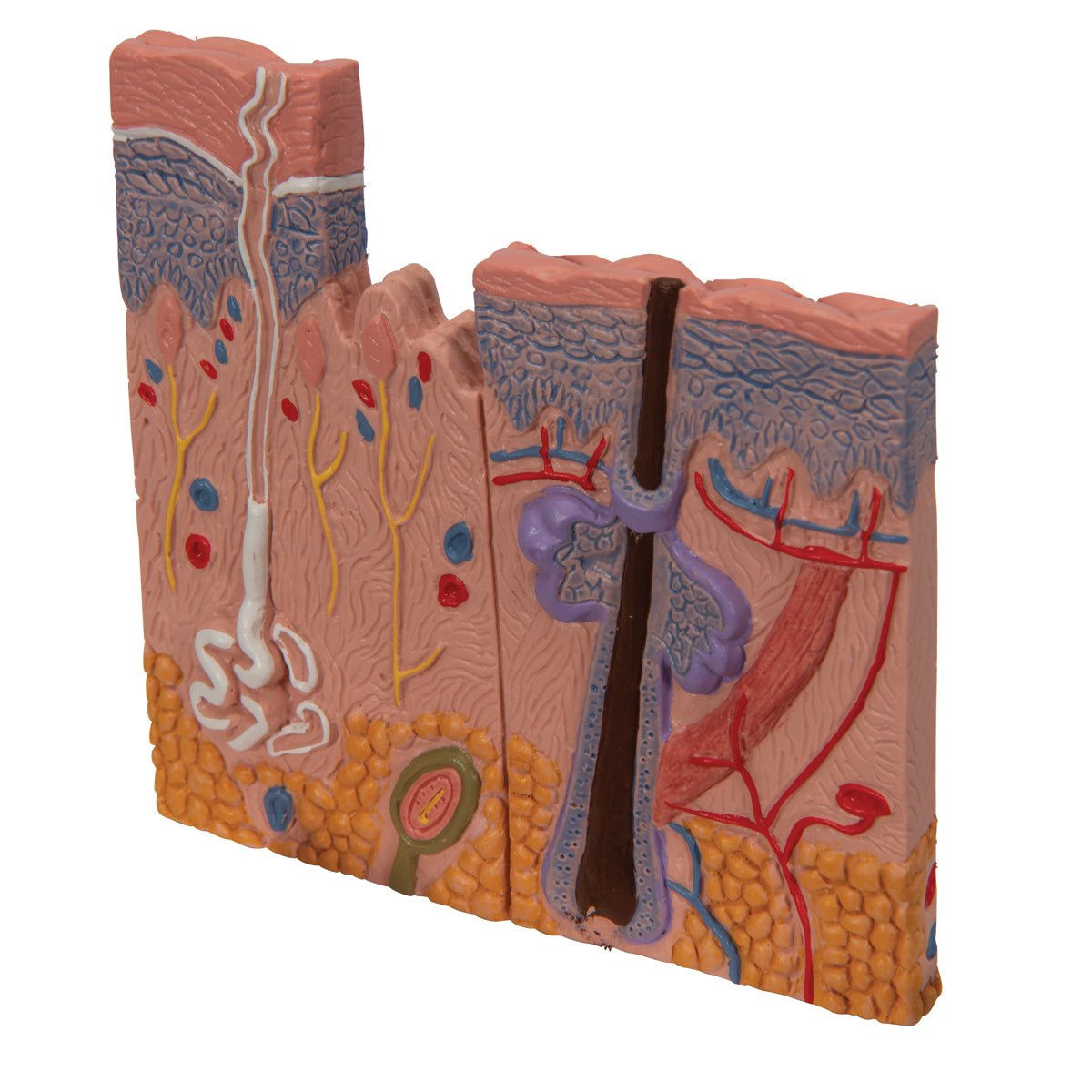
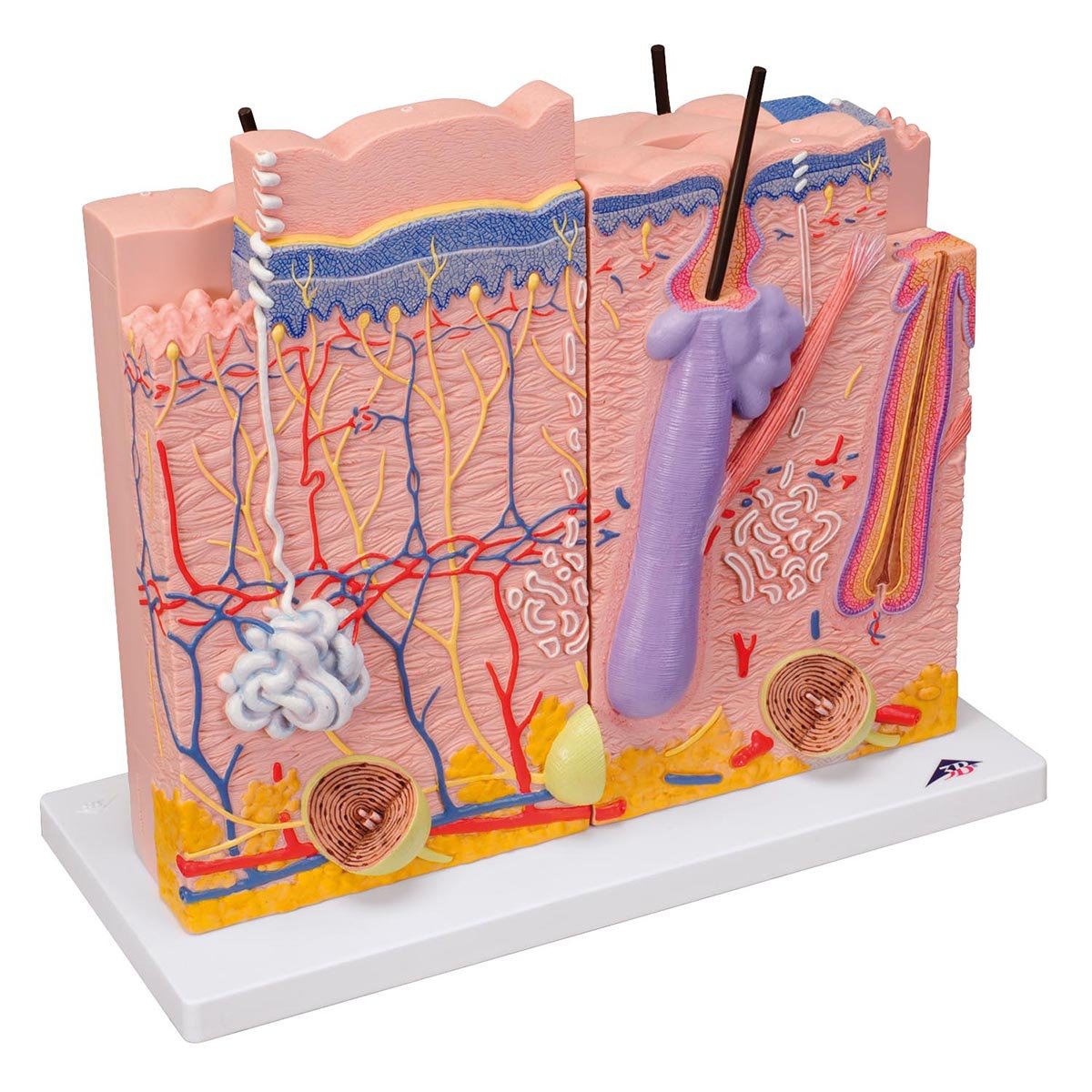
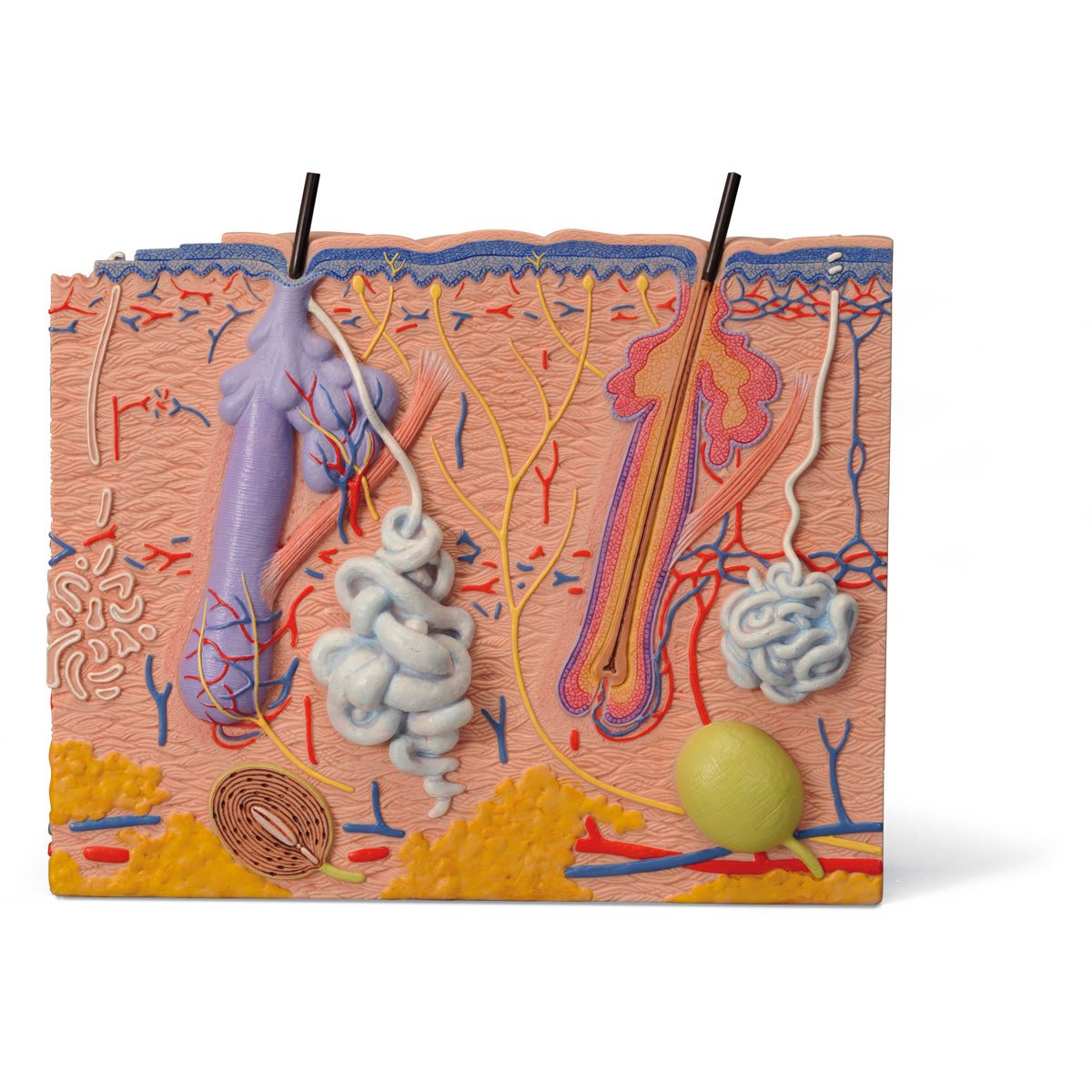
  • Layers of the skin, 70x magnification, anatomical model

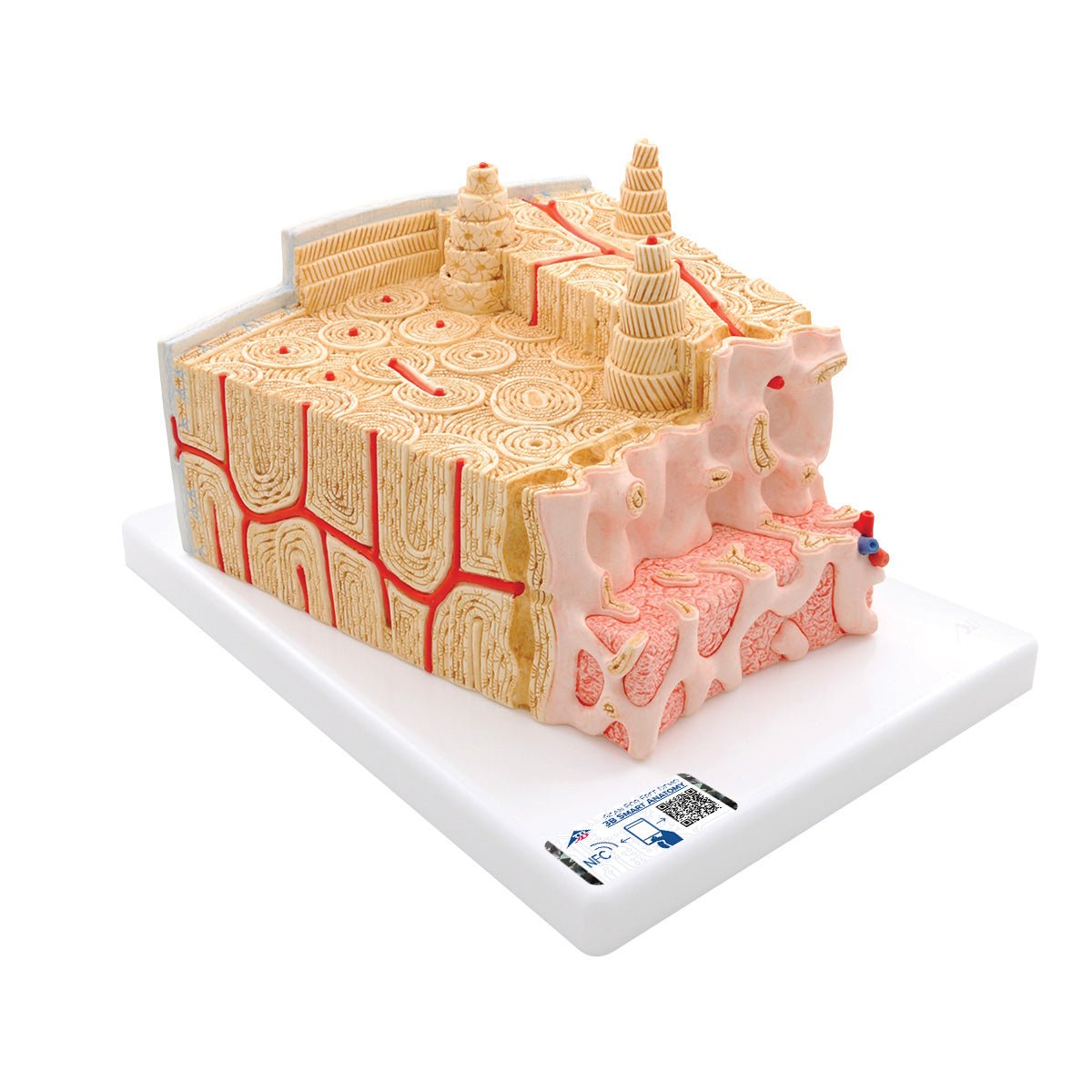
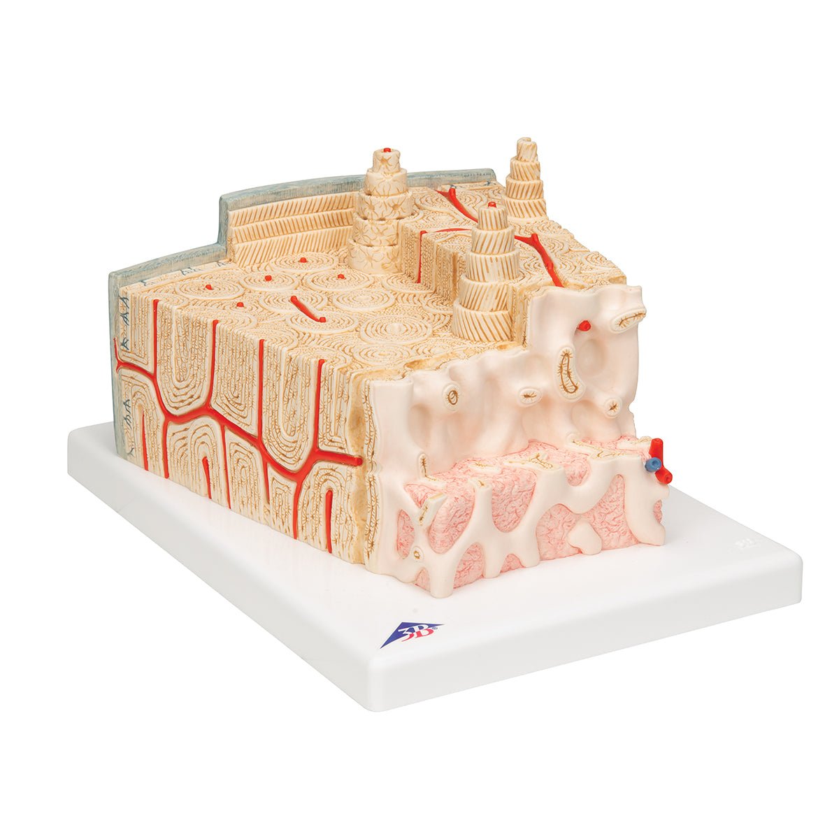
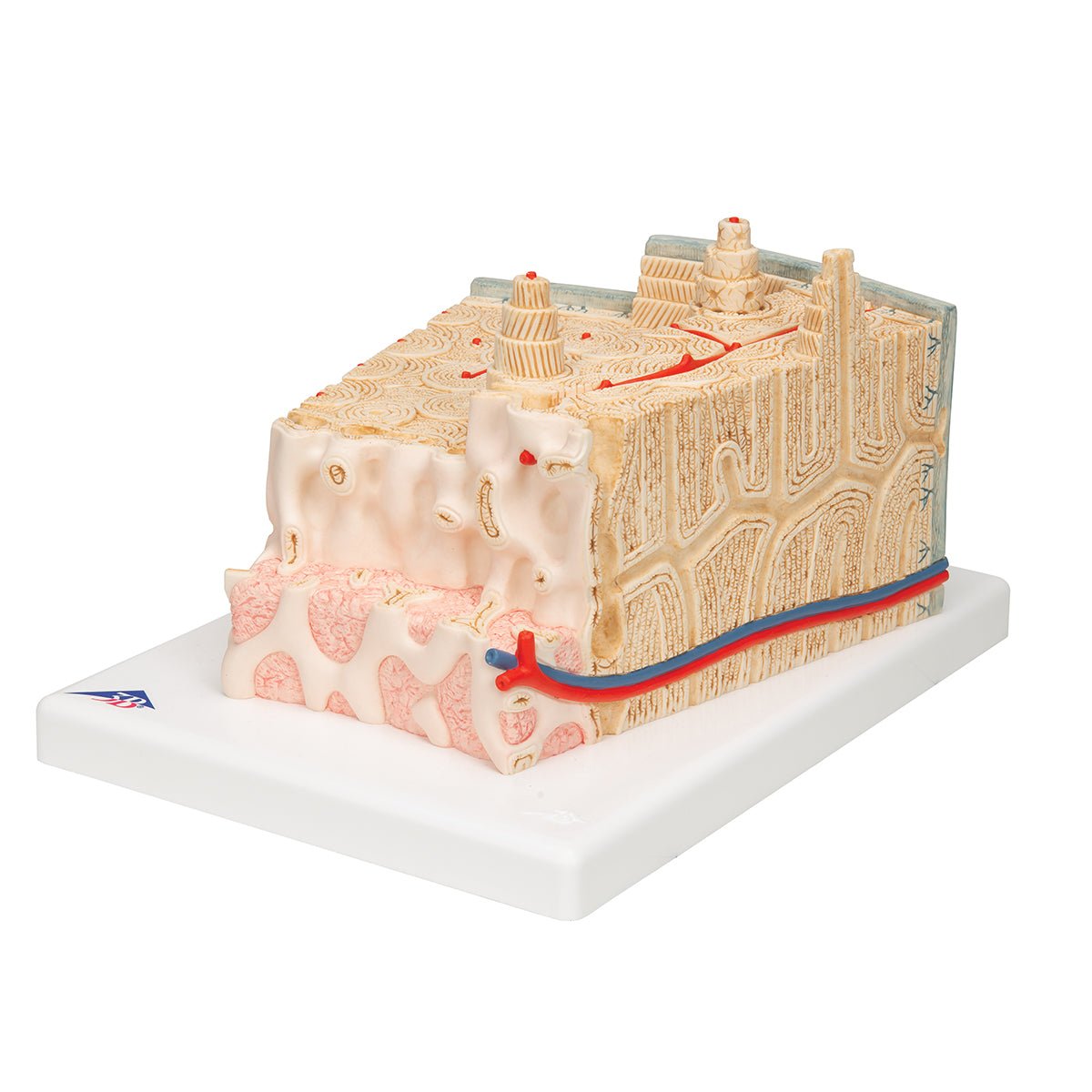
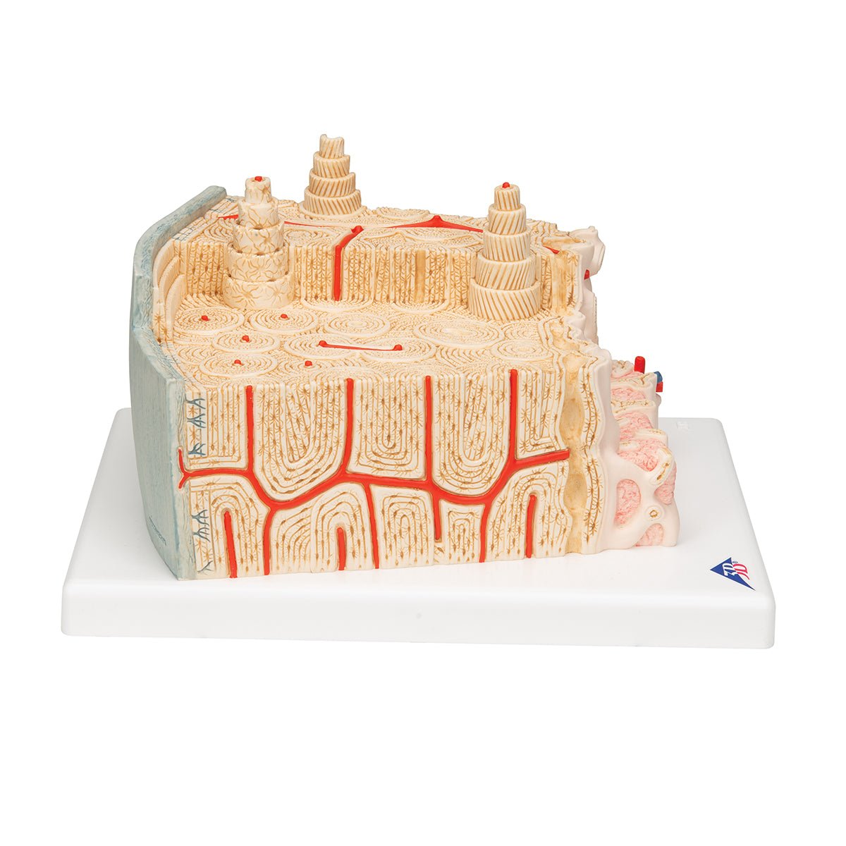
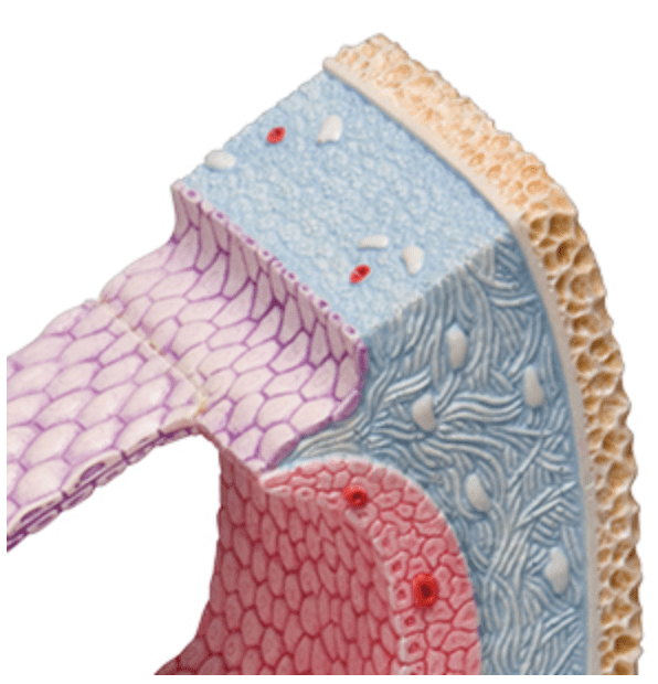
  • Bone tissue including blood vessels, anatomical model, 80x magnification

  • 3 skin areas, 80x magnification, anatomical model

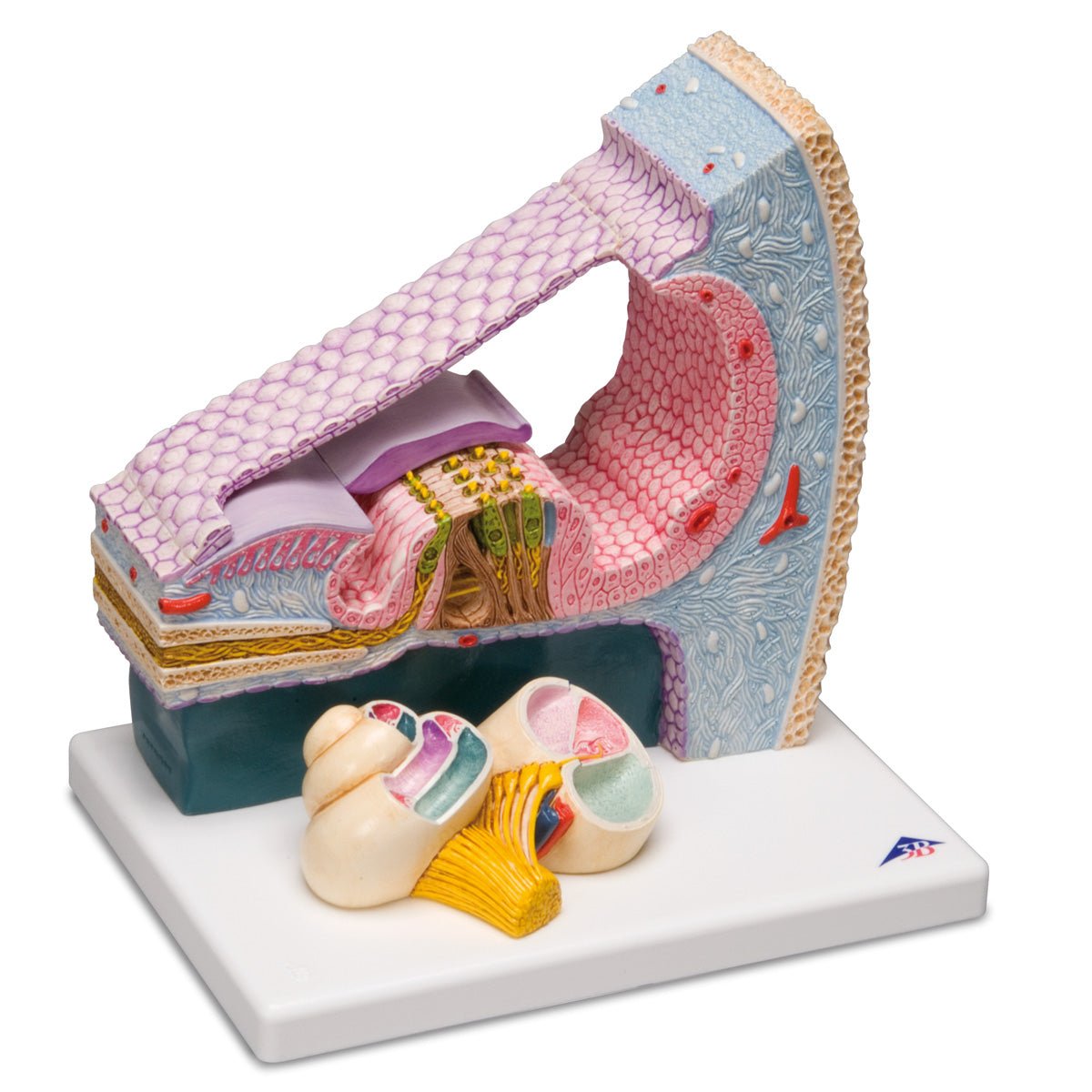
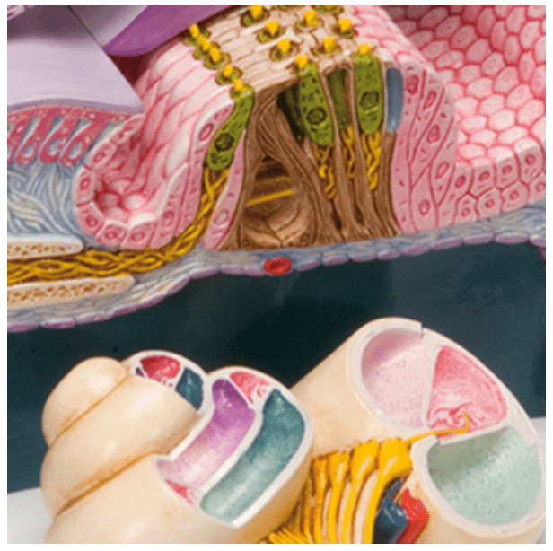
  • Cochlea with cross-section of the ear, anatomical model

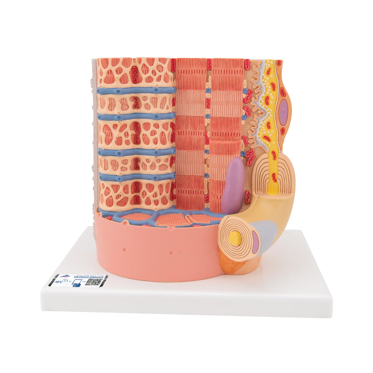
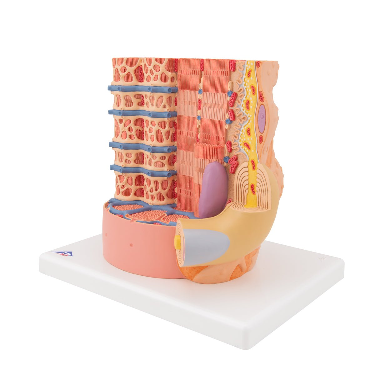
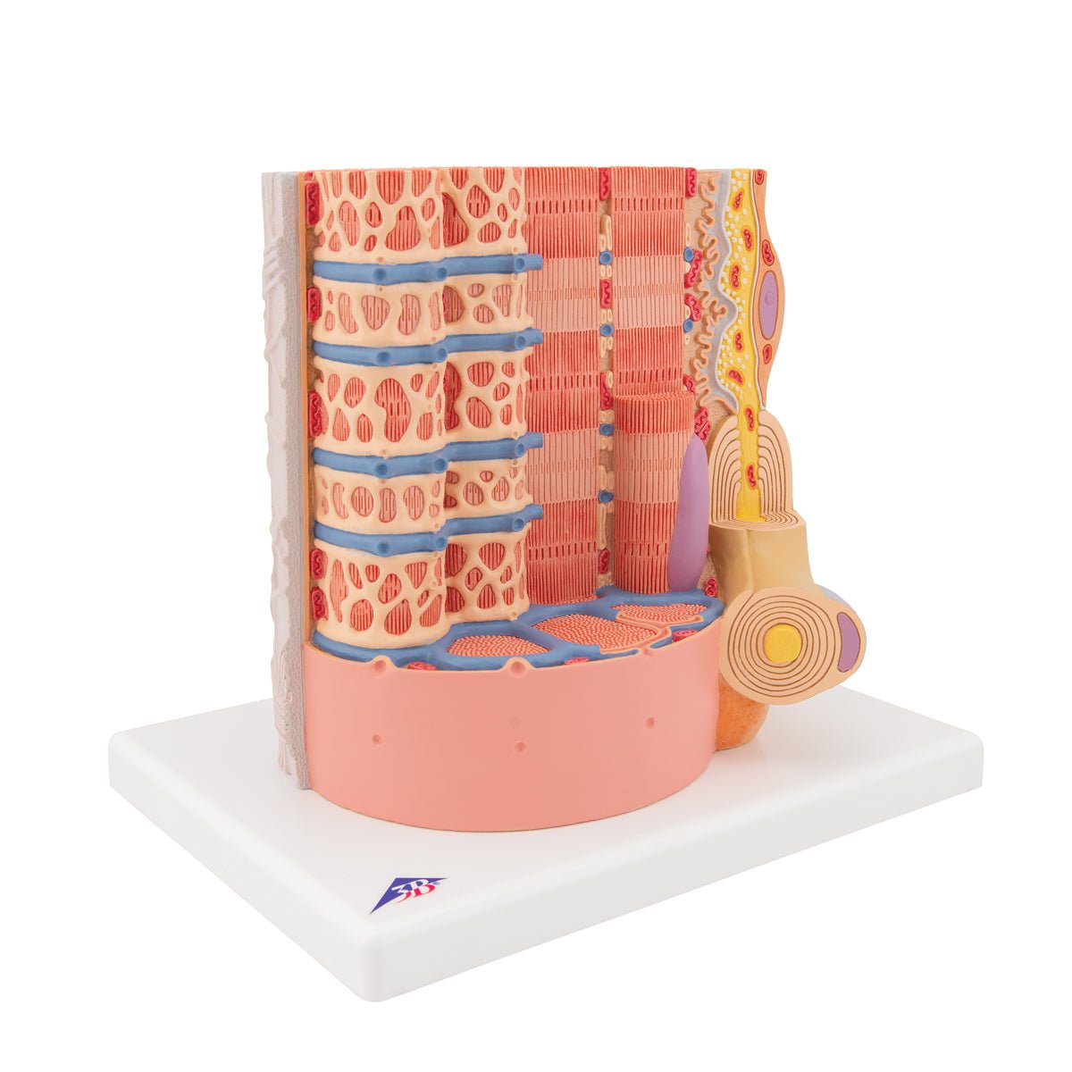
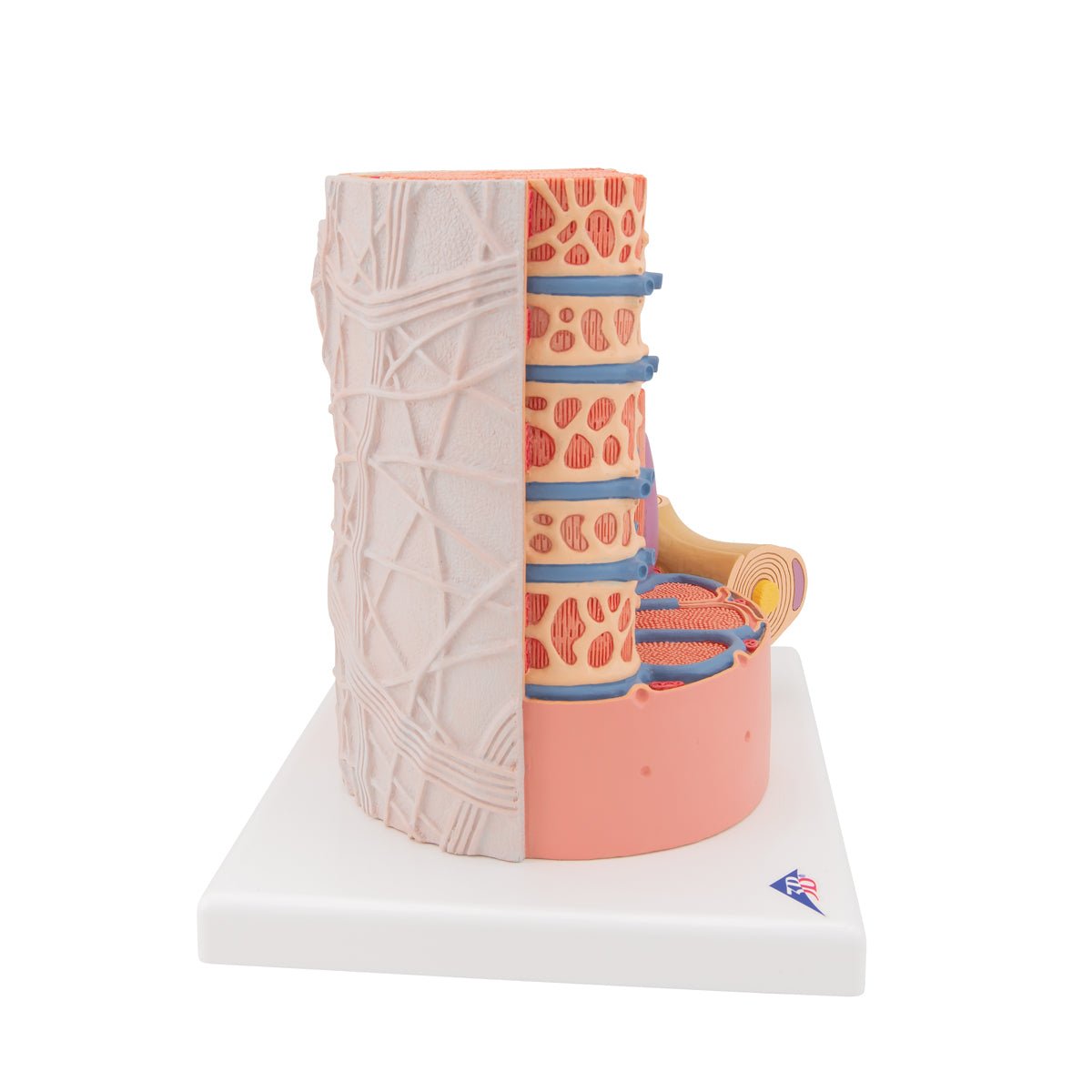
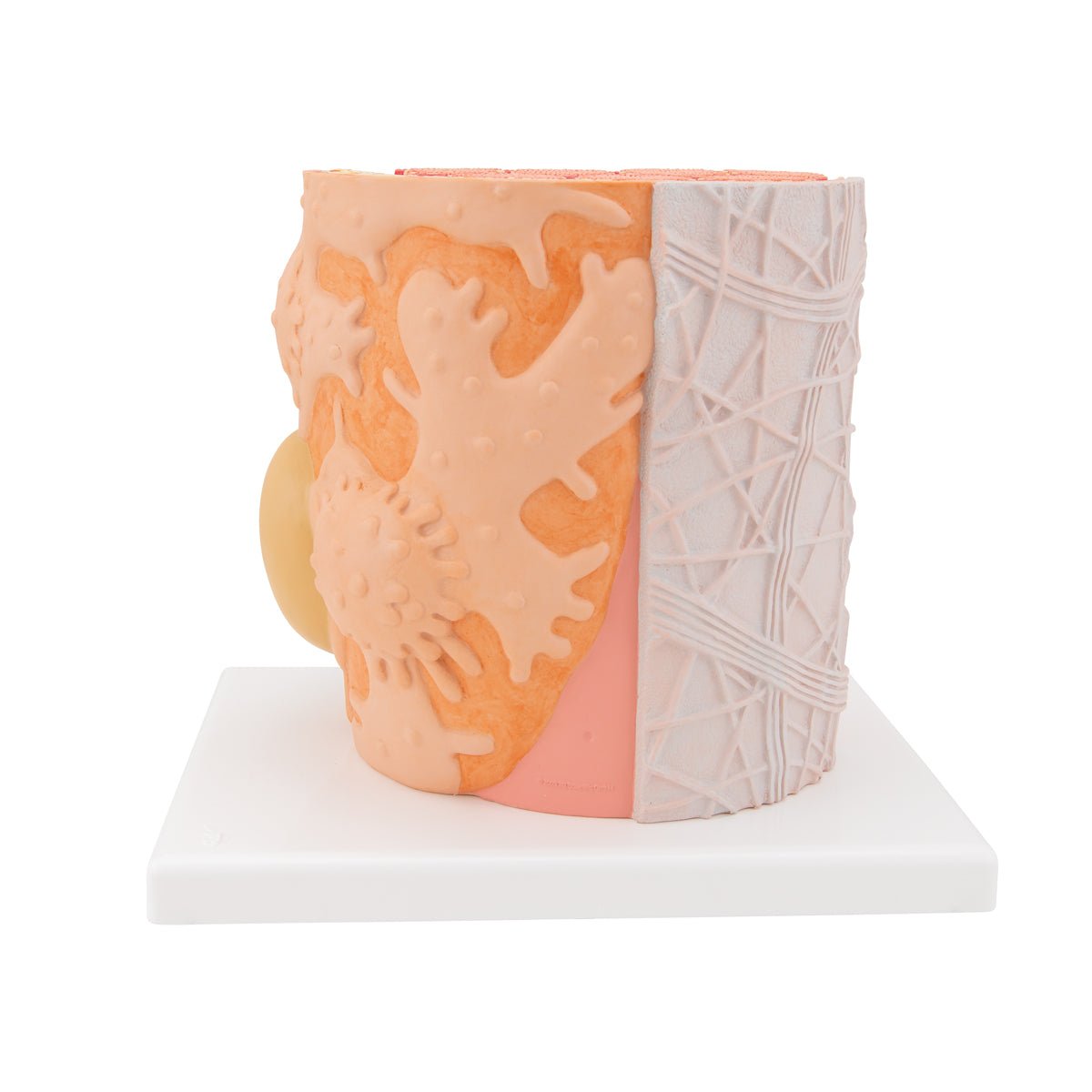
  • Skeletal muscle fiber, anatomical model 10,000x magnified

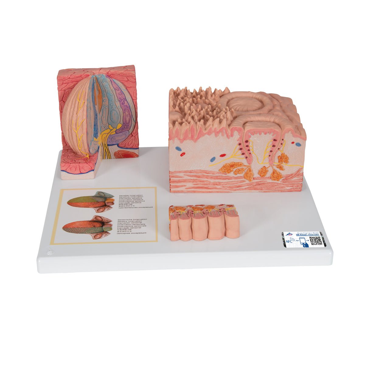
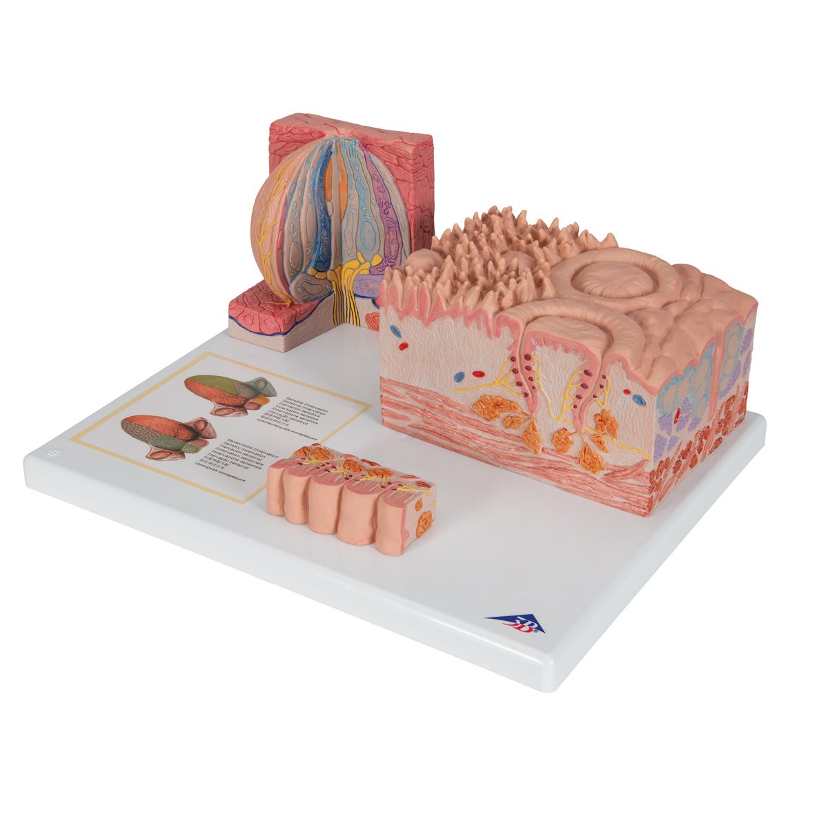
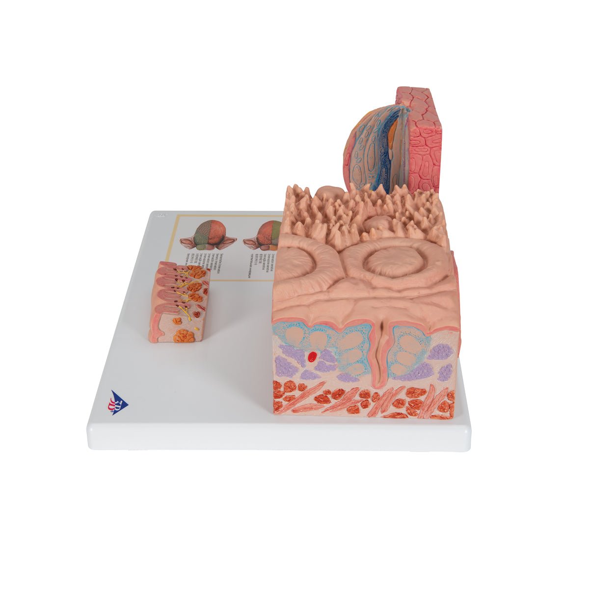
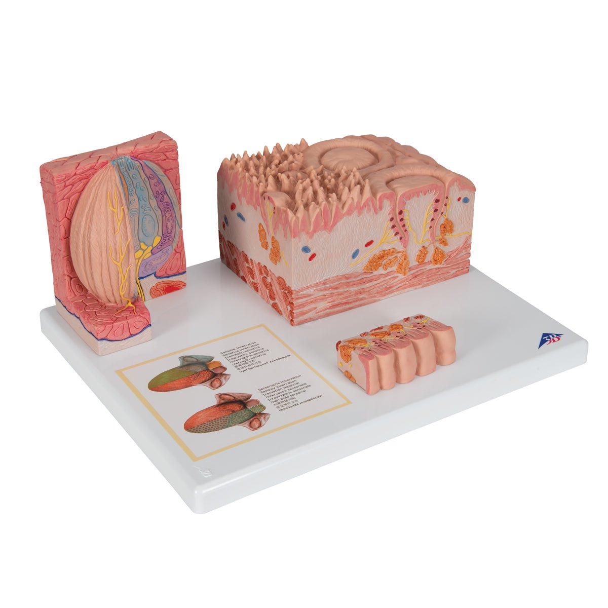
  • Different tissues of the tongue, anatomical model

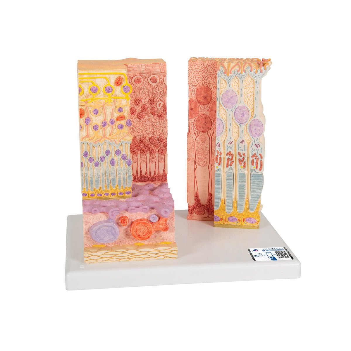
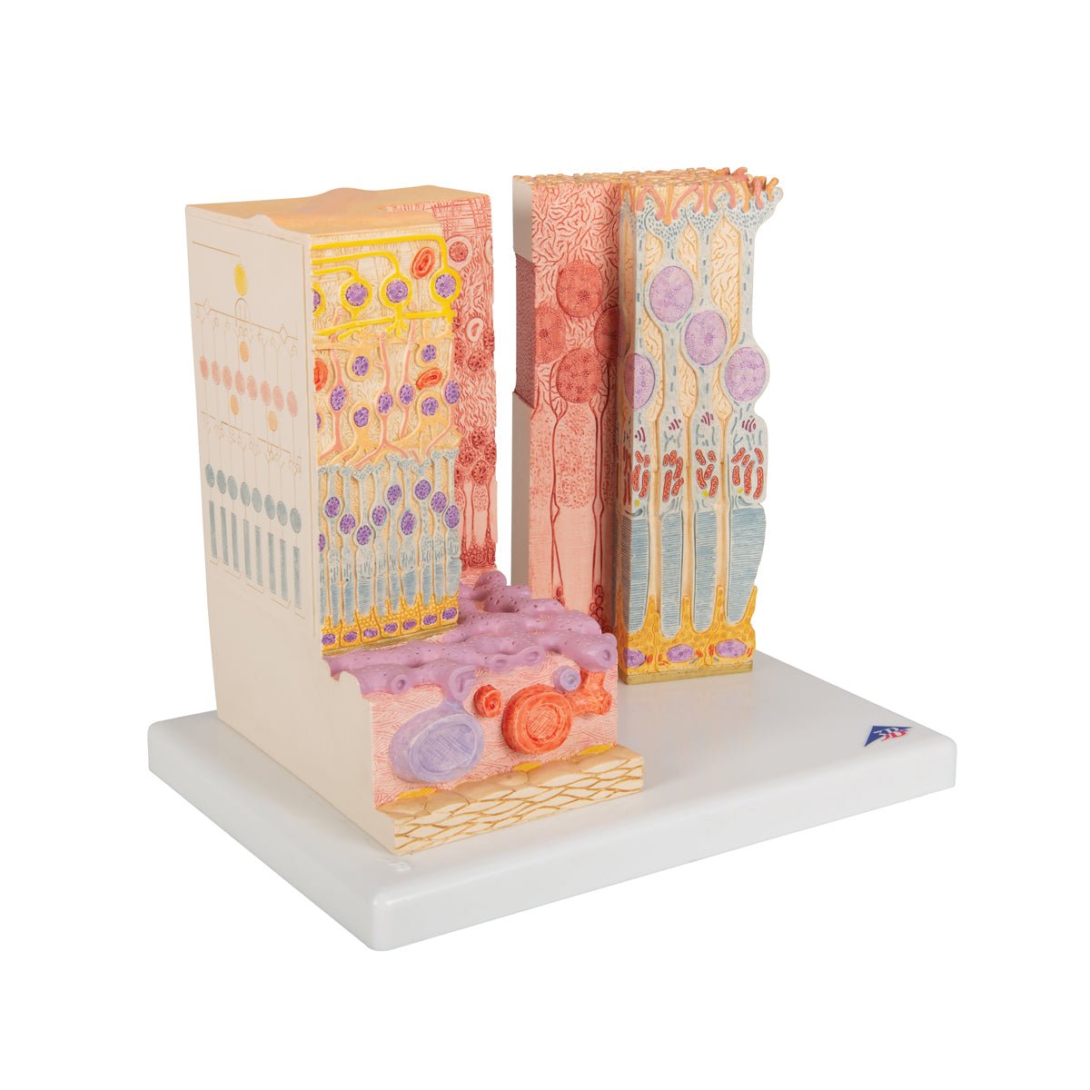
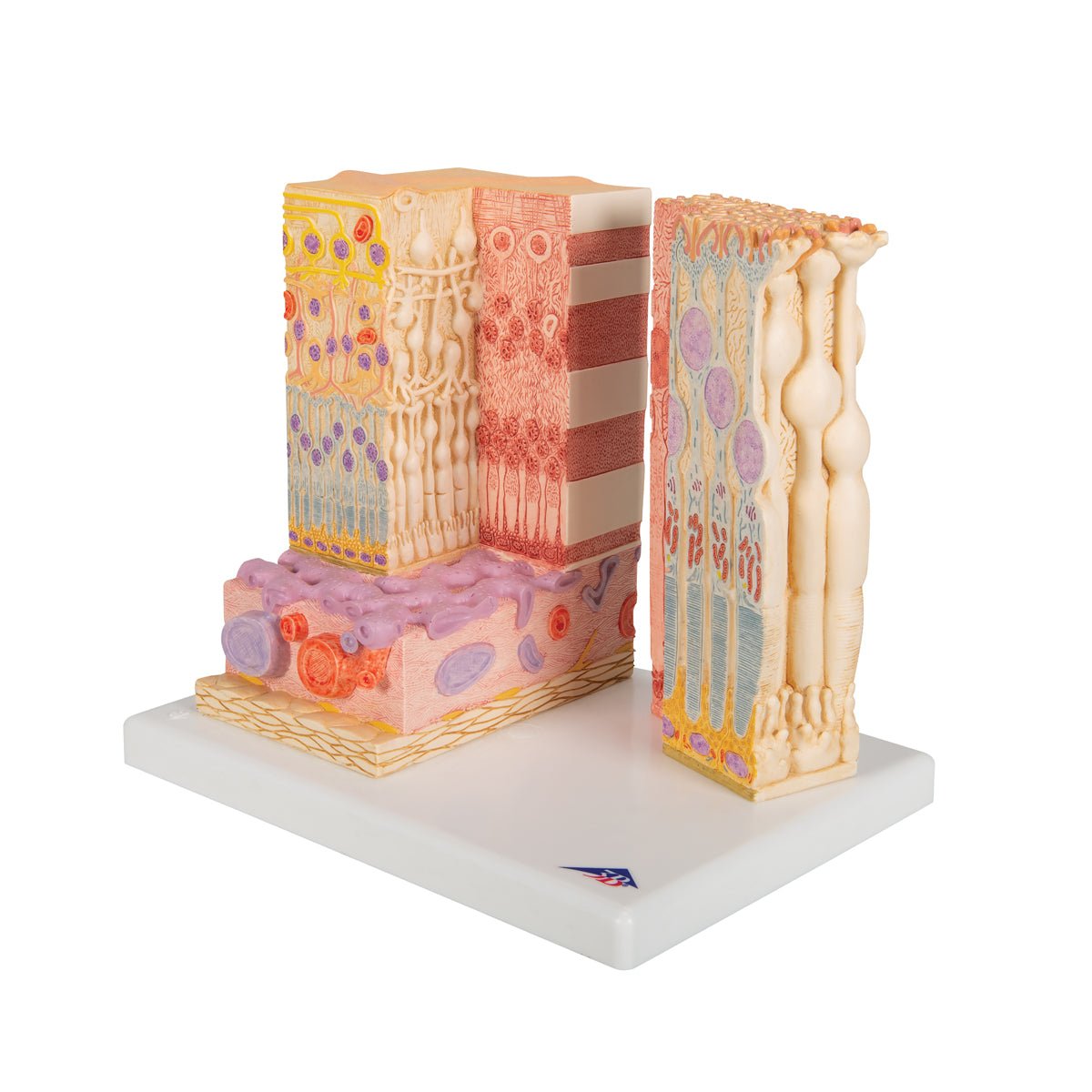
  • Different tissues of the eye, 850x magnified anatomical model

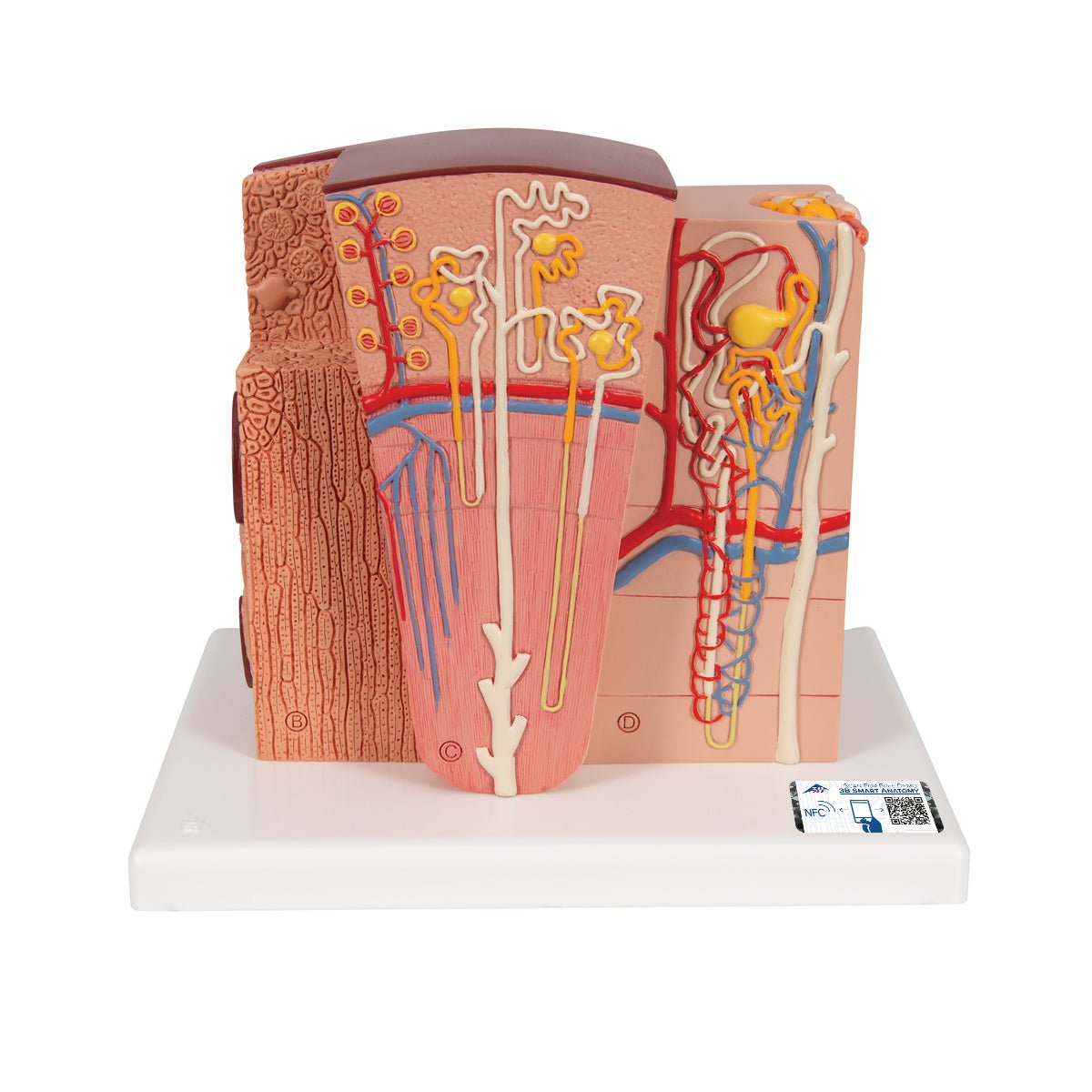
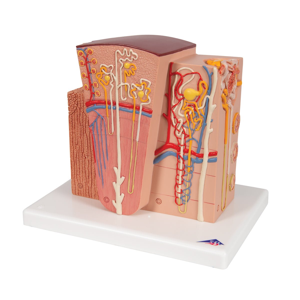
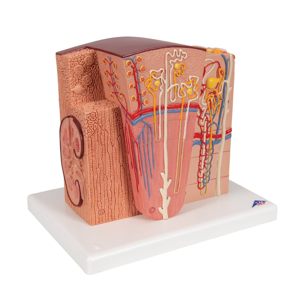
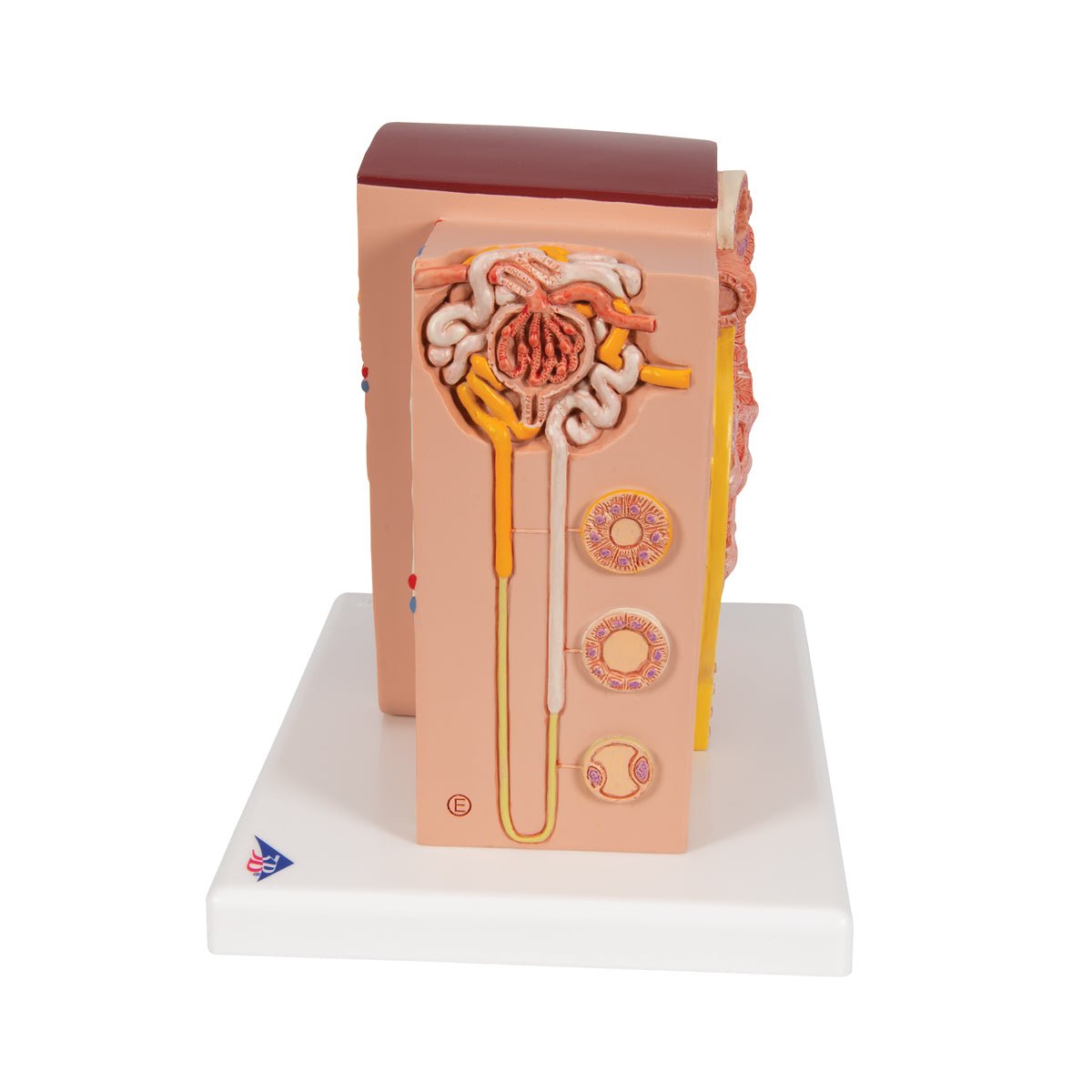
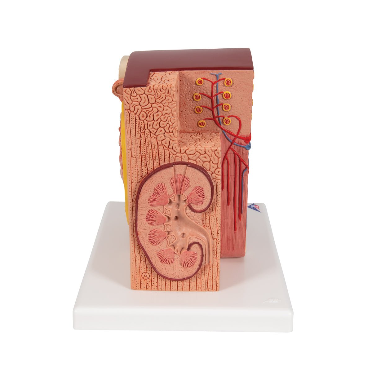
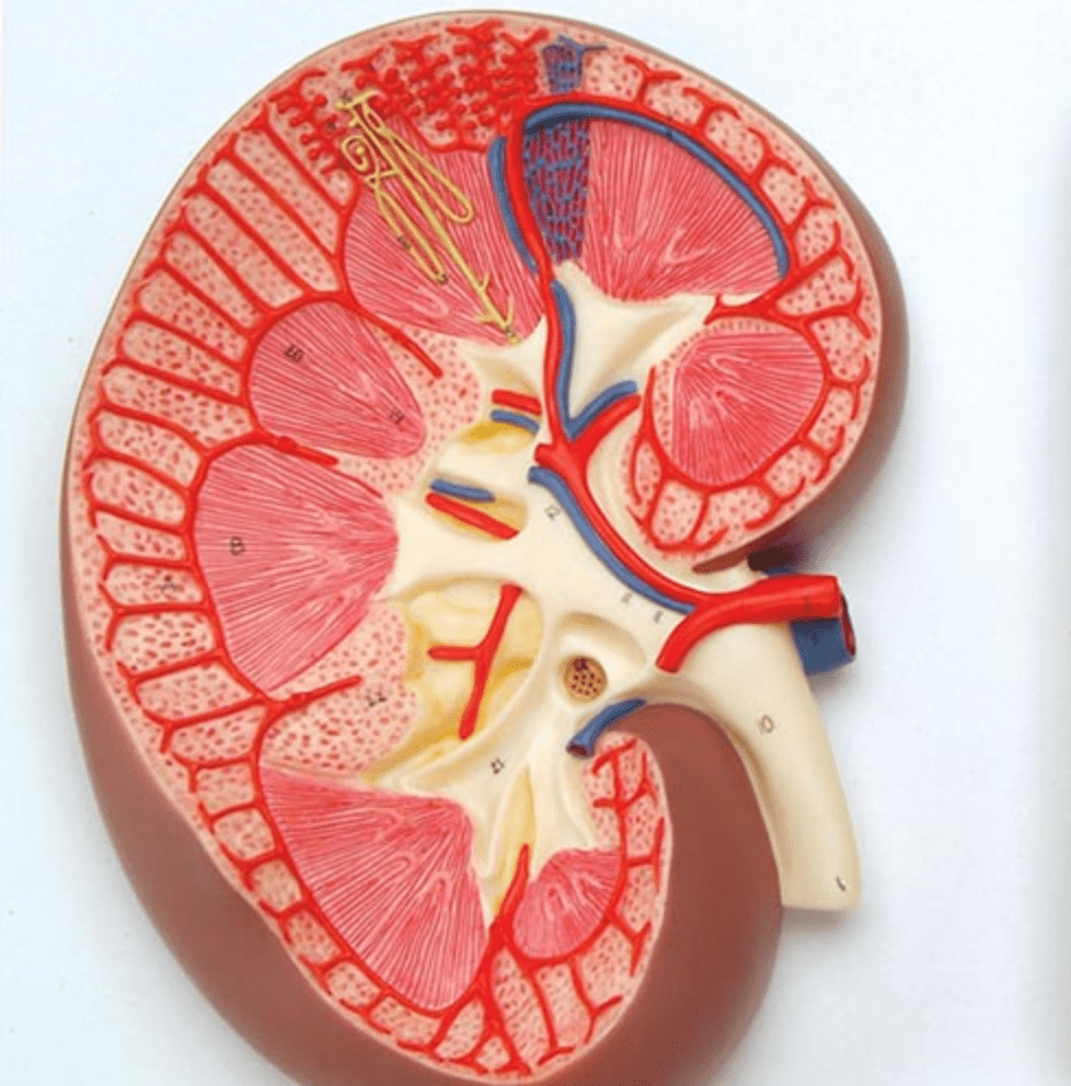
  • Different tissues of the kidney, anatomical model

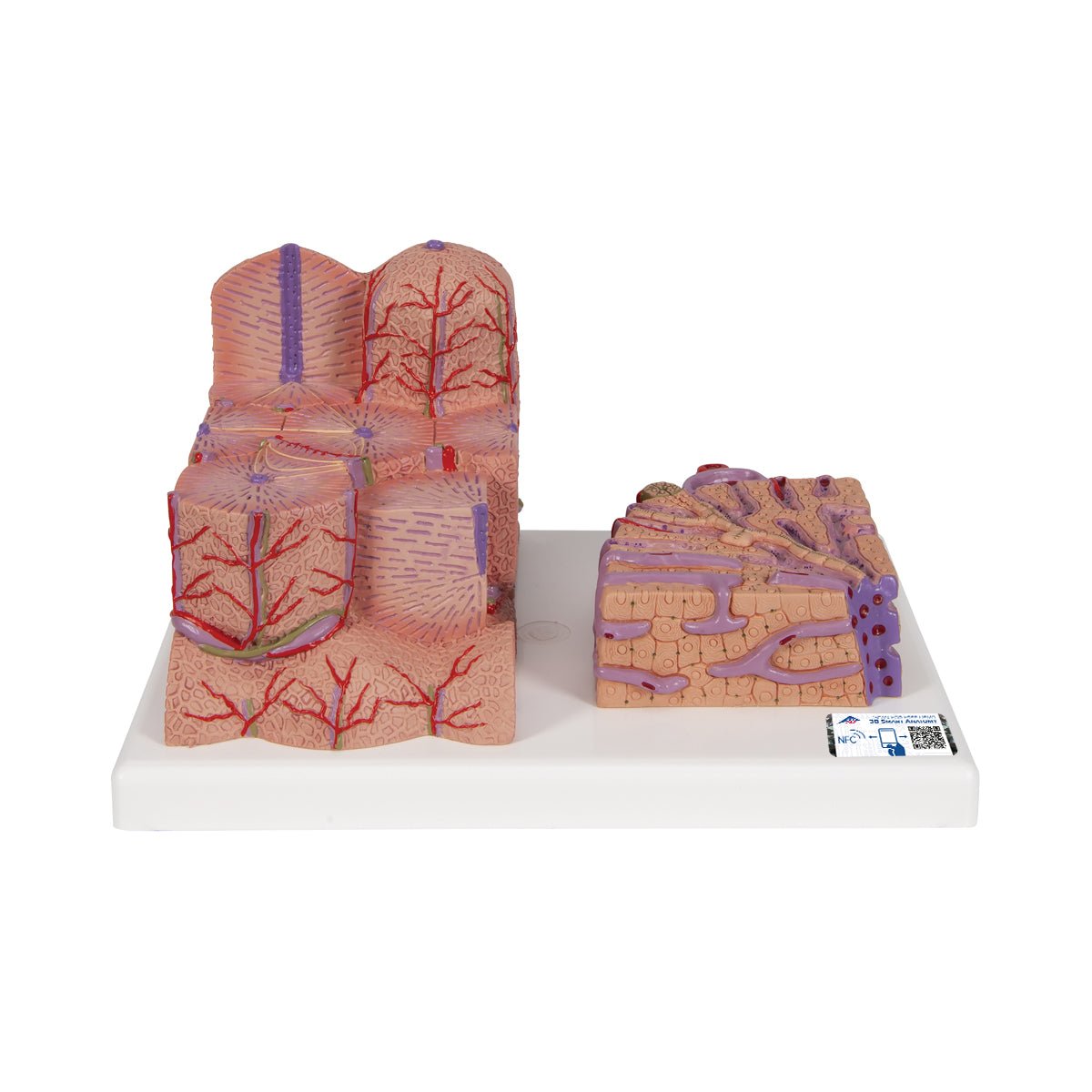
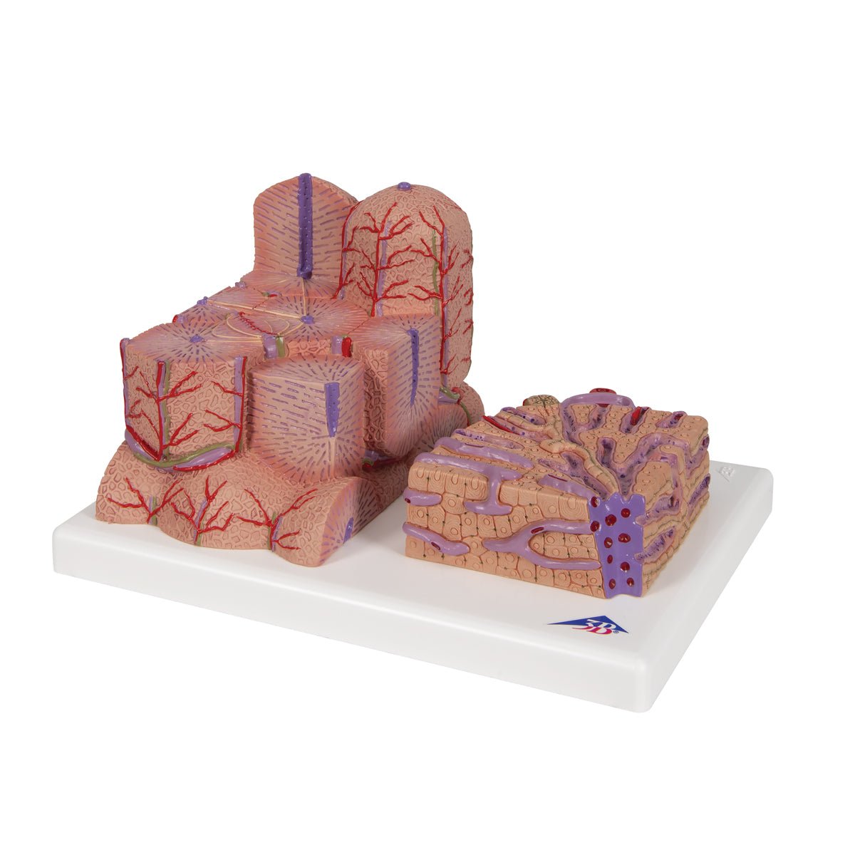
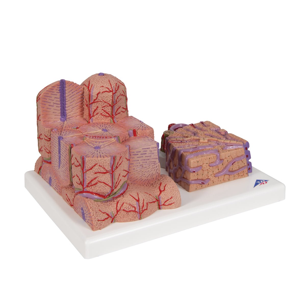
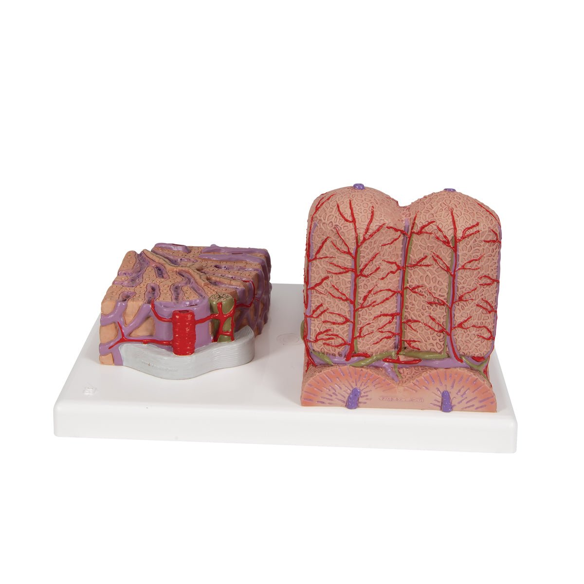
  • Liver tissue with blood vessels, anatomical model 60x magnified

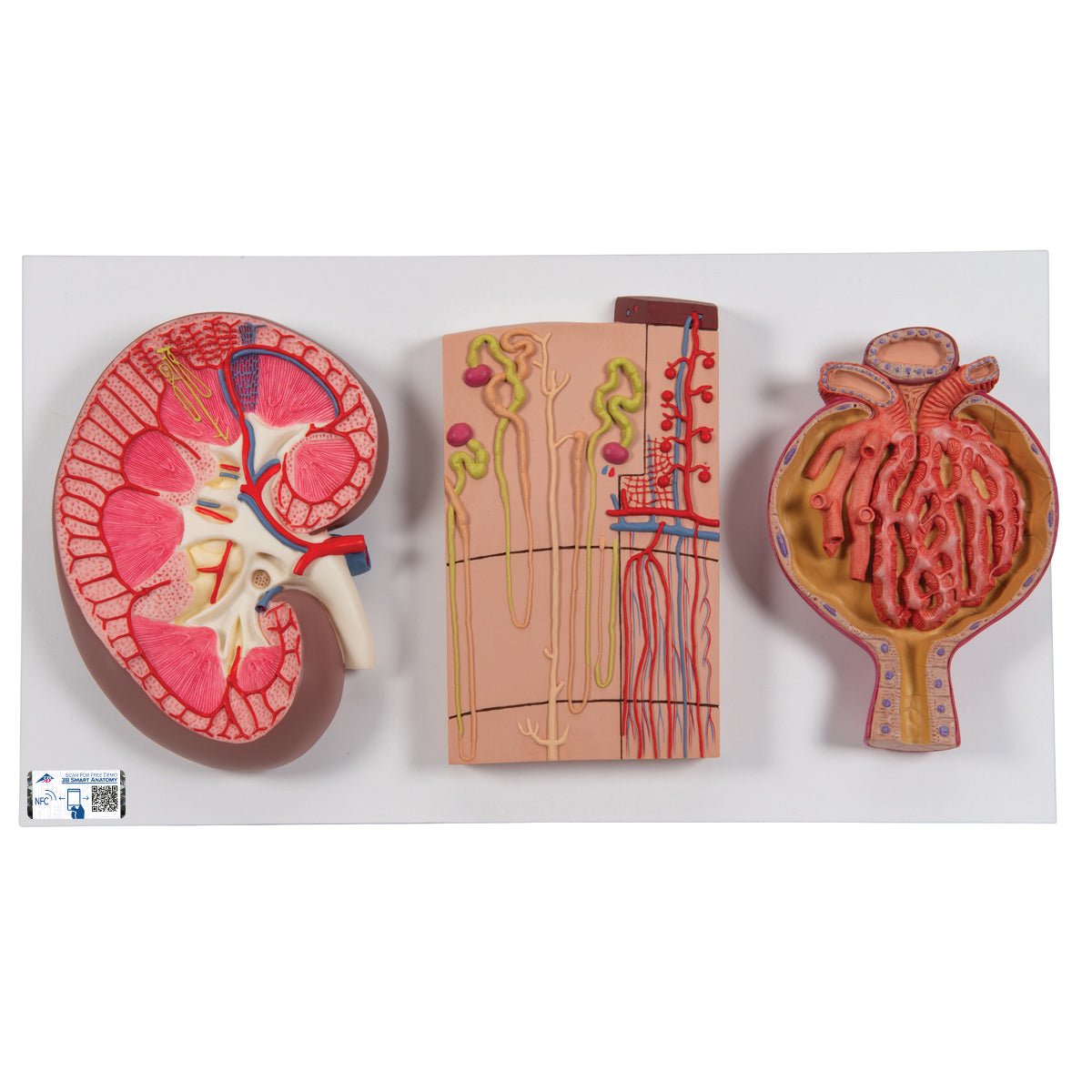
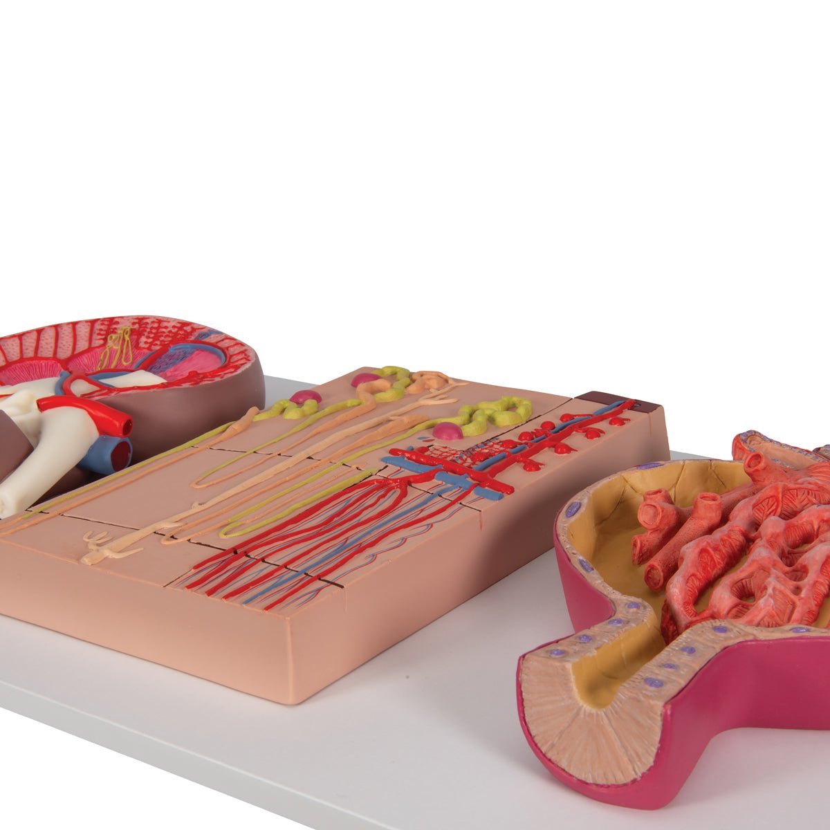
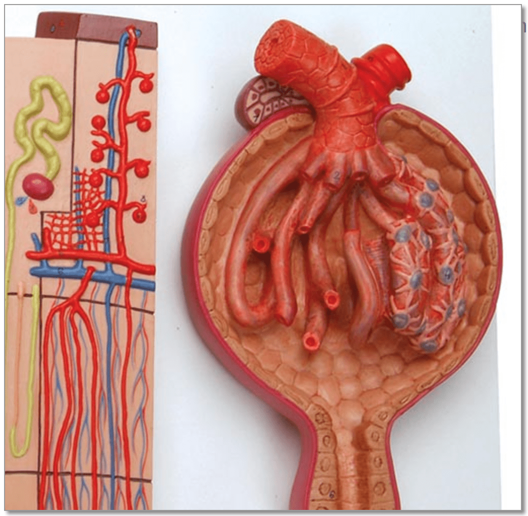
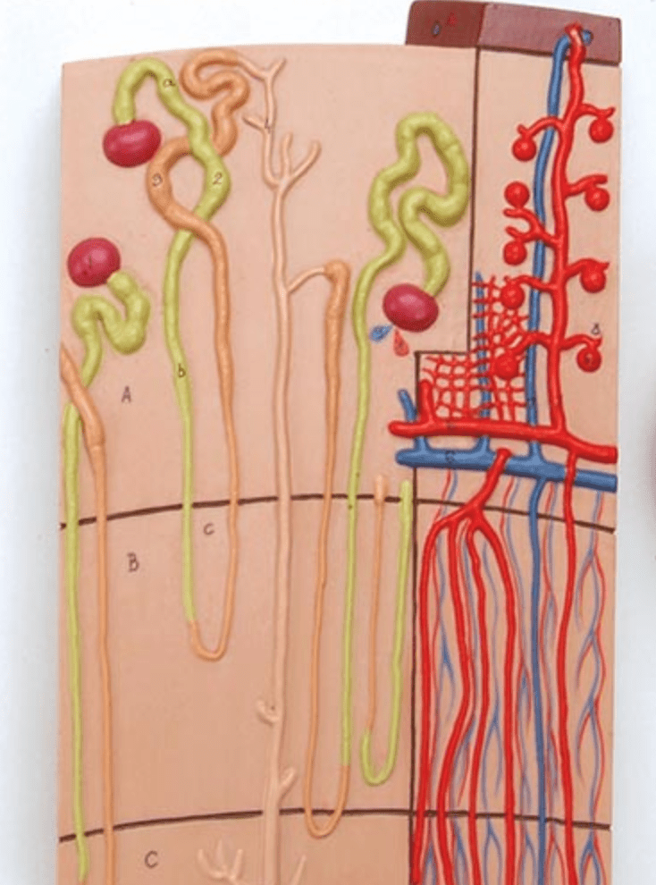
  • Kidney with nephron and renal corpuscle, anatomical model

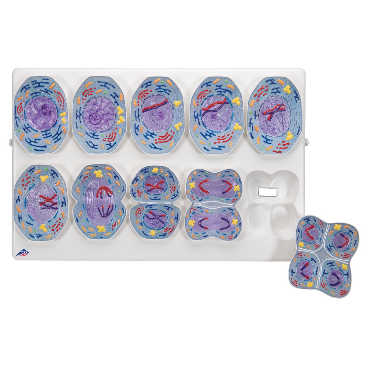
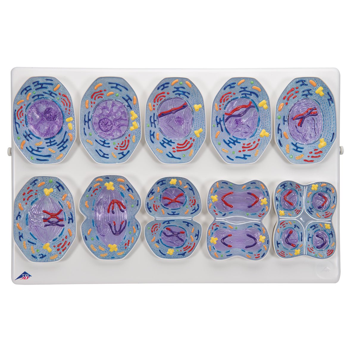
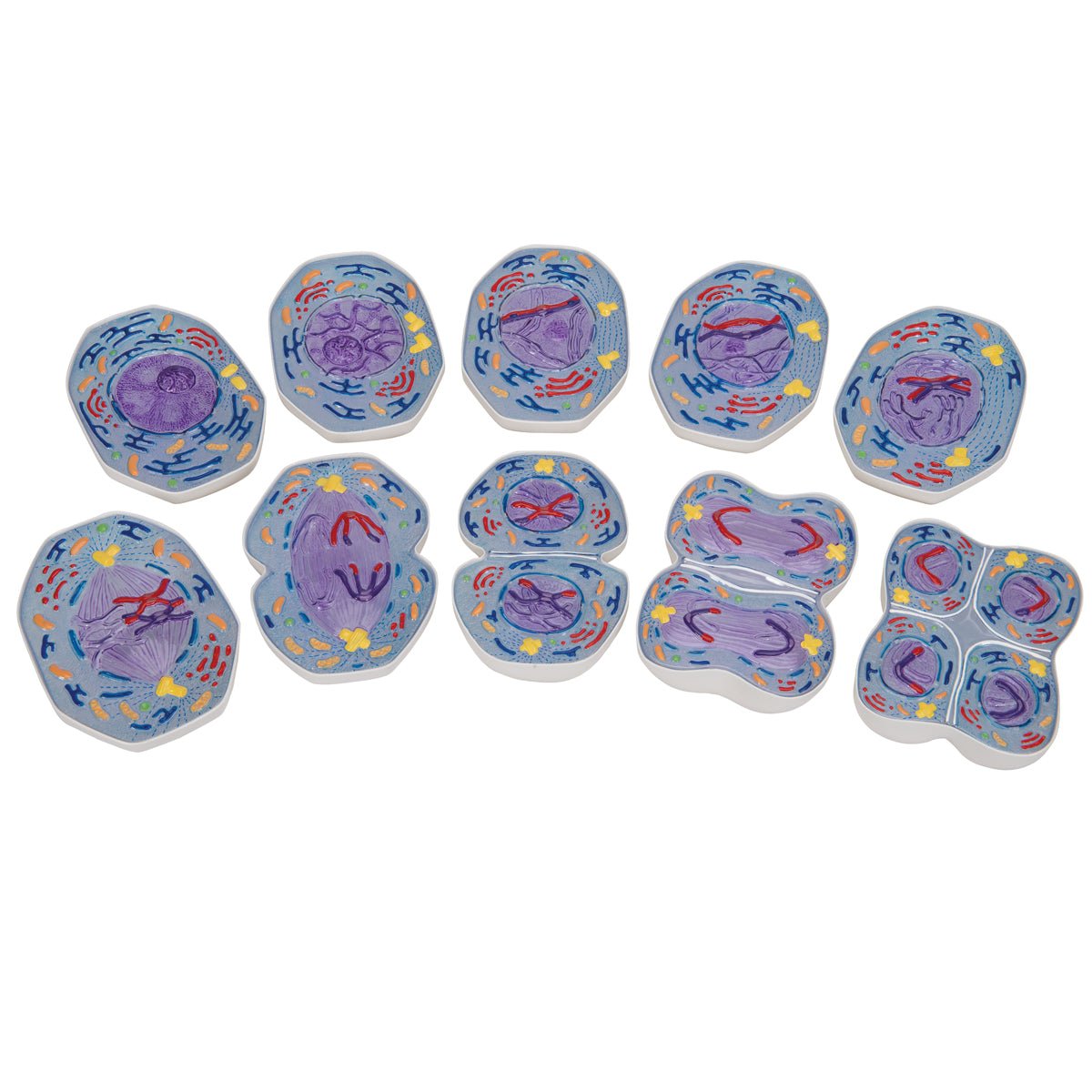
  • Cell division, meiosis, anatomical model

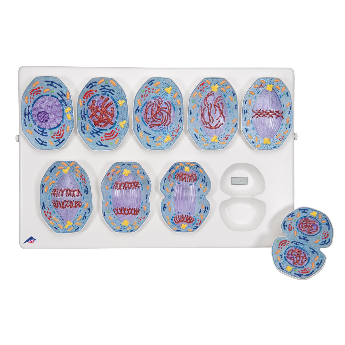
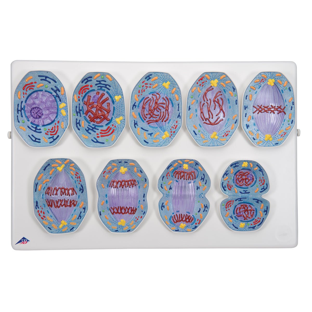
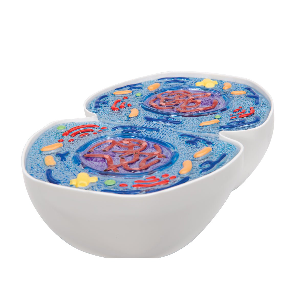
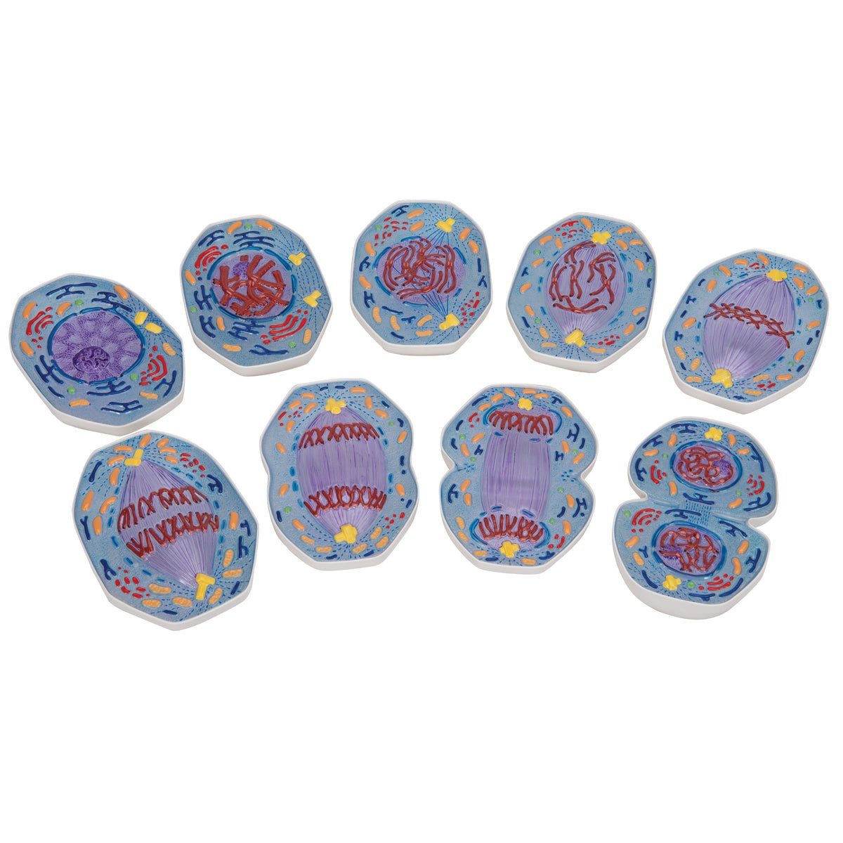
  • Cell division, mitosis, anatomical model

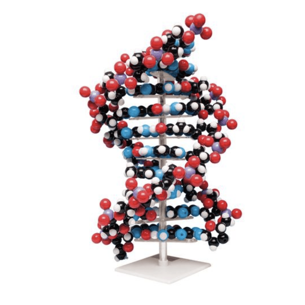
  • DNA strand, 45 cm, anatomical model

  • DNA strand, B shape, anatomical model of 41.5 cm

  • Human cell, 60 cm, anatomical model, 40,000x magnified

Get notified when we add new products

Sign up for our newsletter here