
Imagine being able to look inside the body and experience its internal organs in detail, as if you were there yourself. That is exactly the experience that a model of the body's internal organs can give you.
This article is your complete guide to everything you should know about internal body organ models in 2025. We dive into the function of the models, different types, practical applications, and the latest technological advances.
You will gain insight into how to choose the right model, which criteria are important, and what future prospects are emerging. Whether you are a student, teacher, or worker in the healthcare sector, this guide is tailored to you.
Let's explore together how a model of the body's internal organs can open the door to deeper understanding and better learning.
What is a model of the body's internal organs?
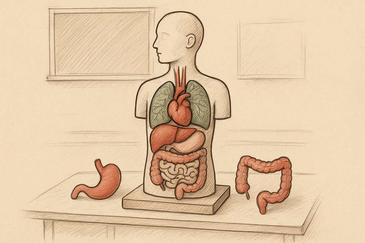
Imagine holding an accurate representation of the human body's internal organs in your hands. A model of the body's internal organs allows you to both see and touch structures that are otherwise only seen in pictures or on screens. Let's dive into what these models actually are and why they are so valuable for both learning and the healthcare sector.
Definition and purpose
A body model is a physical, three-dimensional representation of the organs inside the human body. The purpose of such models is to make anatomy tangible and accessible for teaching, patient communication, research, and even artistic projects. Where posters and digital images only provide a two-dimensional overview, a body model allows the user to see, feel, and explore the location and relationships of organs. In the healthcare sector, models are often used to explain diseases or procedures to patients. According to studies, 82% of medical students use physical models as part of their learning process.
Historical development
The development of models of the body's internal organs began as early as the 19th century, when wax models were used to teach doctors and students. These early models were handmade and often unique, making them both expensive and fragile. With industrialization, it became possible to mass-produce more durable models in materials such as plastic and glass. Over time, technological advances, including 3D printing, have made it possible to produce models with high precision and individualization. Historically, models have played a crucial role in the development of modern medical education and practice.
Types of models
There are many types of models of the body's internal organs, depending on the purpose and the needs of the user. The most common materials are plastic, silicone and modern 3D-printed composites, but glass and even metal are also seen. Some models show the entire body, while others focus on individual organs such as the heart, lungs or liver. Interactive models make it possible to remove organs and examine them individually, while static models are fixed. For example, there are models with removable organs, which facilitate the understanding of complex anatomical relationships.
Pedagogical significance and application
Models of the body's internal organs are of great educational importance, as they make it easier to understand complex anatomical structures. In both the classroom and the clinic, users experience that learning improves significantly when they can work with models. Studies show that people learn up to 70% faster when using 3D models compared to two-dimensional images. Models are increasingly used in universities for practical training and exams. If you want to read more about how students use models in teaching, you can visit For students about anatomical models .
Advantages and disadvantages
A model of the body's internal organs offers several advantages: visual and tactile learning, long durability and realistic reproduction. Models make it easy to explain complex topics to both students and patients. However, they can be expensive and require space for storage, and some materials require careful maintenance. Digital alternatives are more flexible and space-saving, but often lack the physical dimension. User feedback shows that the combination of physical and digital models provides the best learning.
How to Choose the Right Model of Internal Organs
Choosing the right model of the body's internal organs requires careful consideration, whether you are a student, teacher or healthcare professional. To ensure the best result, you should review several criteria that match your needs. Below you will find a thorough guide to help you make the right choice.

Purpose and target group
When choosing a model of the body's internal organs, it is crucial to identify the purpose and target audience. Will the model be used for teaching, patient communication, research, or perhaps as an artistic reference? For example, many educators and medical students choose models that can be disassembled, while doctors often prioritize educational models for patient dialogue.
Examples of usage scenarios include universities, hospitals and private clinics. Statistics show that 60 percent of all models are purchased for educational use. If you have special needs, you can benefit from investigating the possibilities for special solutions and custom work , where models are adapted to specific requirements.
Materials and quality
The choice of material is important for both durability and realism when choosing a model of the body's internal organs. The most common materials are plastic, silicone, glass and 3D printed materials. Plastic models are often robust and affordable, while silicone gives a more lifelike feel.
Choose hypoallergenic and safe materials, especially if the model will be used in education with children or sensitive users. Also consider whether the model should be easy to clean and withstand frequent handling. The quality of the material affects the model's lifespan and daily benefit.
Level of detail and functionality
The level of detail in a model of the body's internal organs is of great importance for learning outcomes. A model with clear structures, color codes and removable organs makes it easier to understand complex anatomical relationships.
Interactive models where you can remove organs and examine them individually are especially popular in education. Some models also show pathological conditions, making them ideal for medical training. Choose the functionality that best supports your learning objective or professional use.
Price and budget considerations
The price of a model of the body's internal organs varies considerably depending on size, level of detail, and brand. Simple models can cost a few hundred kroner, while advanced, interactive models can cost several thousand kroner.
The following table gives an overview of what affects the price:
| Factor | Price influence |
|---|---|
| Size | The bigger, the more expensive |
| Details | More details, higher price |
| Functionality | Interactive ones cost more |
| Mark | Well-known brands are more expensive |
Consider your budget, but don't compromise on necessary features. Tip: Compare multiple suppliers and pay attention to any offers.
Certifications and standards
It is important to ensure that your model of the body's internal organs meets applicable certifications and standards. Especially in the education and healthcare sectors, CE marking and other safety certificates are a requirement.
Reputable manufacturers often document that their models have been tested for quality and safety. Always ask the supplier if you are in doubt about the certifications. This ensures that the model can be used both safely and legally in relevant contexts.
Maintenance and storage
Proper maintenance will extend the life of your internal organs model. Clean the model regularly with mild detergents, avoiding harsh chemicals. Store the model dry and out of direct sunlight to prevent discoloration and material degradation.
Mistakes like dropping the model or exposing it to moisture can significantly shorten its lifespan. Use storage boxes or cabinets if the model is not on display. A good maintenance routine will ensure that your investment lasts for many years.
Technological Advances in Anatomical Models 2025
Technological advances have transformed how we work with a model of the body's internal organs. In 2025, we will see a significant evolution where innovation and precision go hand in hand. Below, we explore the key trends shaping the models of the future.
3D printing and digitization
3D printing has revolutionized the production of a model of the body's internal organs. Today's 3D printed models offer unprecedented precision and customization. University hospitals can now order patient-specific models, improving both planning and training.
Tailor-made solutions mean that models can be made according to individual needs. Statistics show that 35% of new models are now produced with 3D printing. This development has made it easier to visualize complex structures and diseases. Digitalization also contributes to faster production and lower costs.
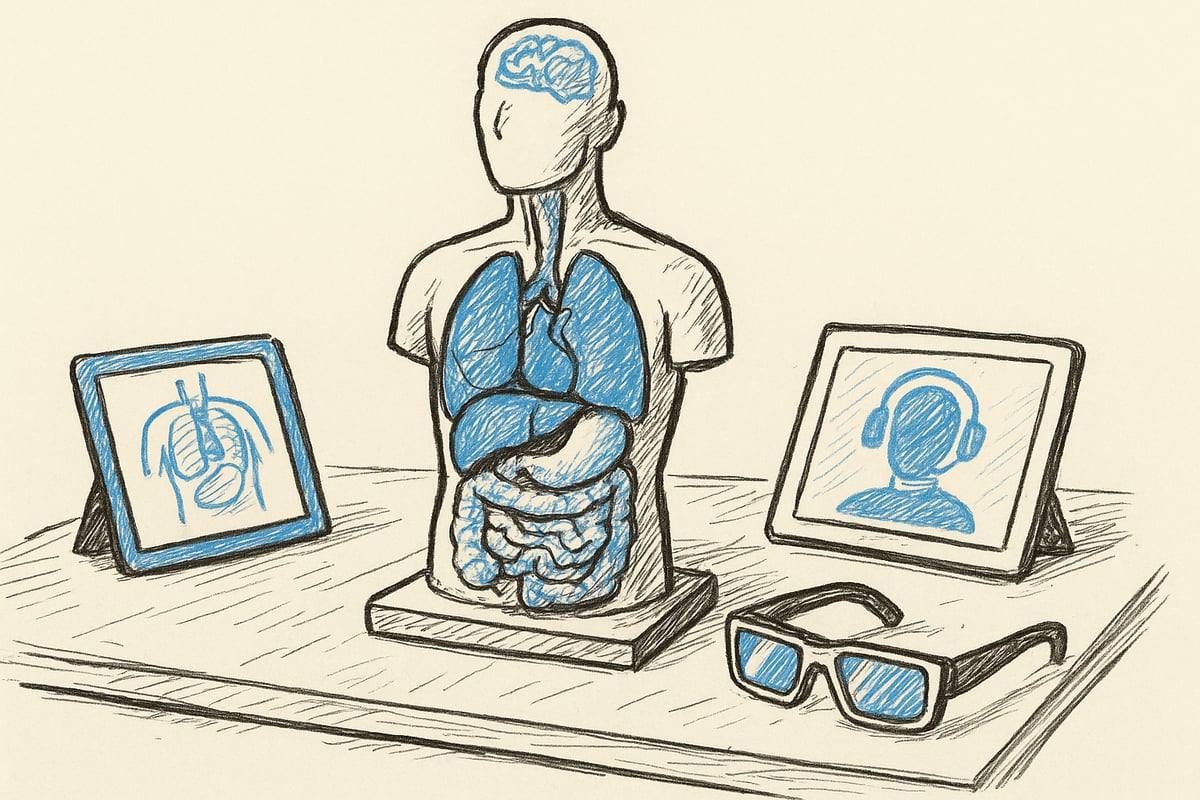
Interactive and augmented reality solutions
Today, physical models are often combined with digital technologies such as augmented reality (AR) and virtual apps. When a model of the body's internal organs is supplemented with AR, the user can experience layered information directly on the model. This provides a more interactive and engaging learning experience.
For example, AR apps can show blood flow or disease progression in real time. Many educators also integrate anatomical posters and visual aids to further reinforce understanding and create coherence between physical and digital resources. This combination significantly increases the learning effect.
Sustainability and materials
Sustainability has become a key factor in the development of a model of the body's internal organs. Manufacturers are now using bioplastics and recyclable materials to reduce their environmental impact. This shift has resulted in a 20% reduction in the carbon footprint of new models.
Environmental concerns are also reflected in the choice of packaging and distribution methods. Schools and hospitals are increasingly demanding models that meet green standards. The future points towards even more environmentally friendly solutions, where both product and production are well thought out.
Customization and individualization
The ability to customize a model of the body's internal organs to meet specific needs is greater than ever before. Patient-specific models are now routinely used in surgical planning, increasing safety and precision during procedures.
The teaching also features tailored models that match the curriculum or specific learning goals. This individualization makes it possible to include rare diseases or unique anatomical variations in the training. The result is a more targeted and effective learning environment.
Future trends
The future of a model of the body's internal organs holds even more technological advances. Integration with digital learning platforms is expected to grow, with hybrid solutions combining the best of both worlds. Experts predict that models will become even more interactive and intelligent.
Also expect an increase in the use of artificial intelligence to create self-explanatory models that adapt to the user's level. These trends point towards a learning environment where anatomy education becomes more intuitive, accessible, and engaging for all users.
Practical Applications: From Teaching to Patient Communication
Models of the body's internal organs have taken on a central role in many practical contexts. From educational institutions to hospitals and public events, these models are used to make complex anatomy understandable to both professionals and laypeople. The visual and tactile elements create a unique learning environment where theory and practice meet. Below, we review six important areas where a model of the body's internal organs creates value.
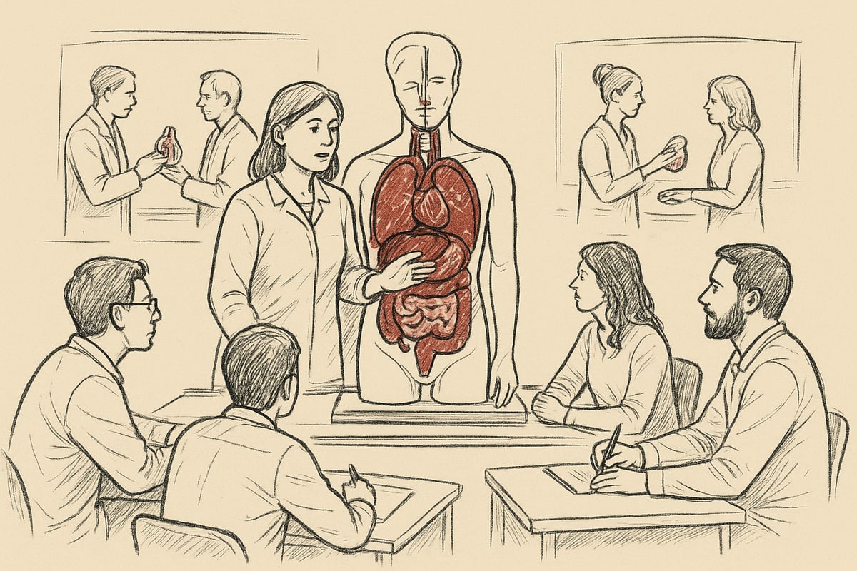
Use in education and exams
A model of the body's internal organs is indispensable in modern education at all levels. Medical, nursing and biology students are given a concrete tool that strengthens their understanding of anatomy. For example, models are used in OSCE exams, where students must demonstrate practical skills and anatomical knowledge.
According to studies, 90 percent of medical faculties use physical models in teaching. This is because a model of the body's internal organs makes it easier to visualize complex structures. Many teachers find that students learn faster when working with physical models. Read more about this in Anatomical Models in Teaching , which describes the importance of models for learning.
Compared to digital alternatives, the models provide a direct, sensory experience that supports both memorization and practical training.
Patient guidance and communication
In clinical consultations, a model of the body's internal organs can be crucial for patient understanding. When the doctor uses a physical model to explain diseases or treatments, patients experience greater comfort and insight.
In oncology departments, the use of models has been shown to significantly improve dialogue. Patients can ask more precise questions because they can visually follow the doctor's explanations. This often leads to better treatment outcomes and higher patient satisfaction.
A model of the body's internal organs makes it possible to show exactly where a disease is located or how an operation will take place. It makes the conversation more concrete and understandable for everyone involved.
Research and development
In medical research, a model of the body's internal organs plays an important role. Scientists use models to test surgical procedures, develop new techniques, and simulate interventions without risk to patients.
By using a model of the body's internal organs, researchers can save both time and resources. It is possible to repeat complicated procedures multiple times, which increases the quality of research results. The models can also be used to demonstrate new discoveries to colleagues and students.
In development work, the models provide an opportunity to refine methods before they are implemented clinically. This contributes to increased patient safety and innovation in the healthcare sector.
Art and visual communication
A model of the body's internal organs is not just reserved for education and healthcare. Many artists use anatomical models as inspiration or as part of their works and exhibitions.
Galleries and museums often feature installations where a model of the body's internal organs forms the framework for visual interpretations of the human body. These projects bridge the gap between science and art and make anatomy accessible to a wider audience.
The models are also used in visual communication, for example to illustrate articles, books or online media. This creates a deeper understanding and fascination for the structure of the body.
Training of healthcare professionals
Simulation training using a model of the body's internal organs has become standard practice in many nursing schools and hospitals, allowing staff to practice realistic scenarios such as emergency care or surgical procedures.
About 75 percent of nursing schools use advanced models in their teaching, making it possible to train practical skills in a safe environment where mistakes do not have consequences for real patients.
A model of the body's internal organs can be equipped with moving parts and replaceable organs to make training as realistic as possible. This strengthens staff's confidence and skills when they encounter patients in real life.
Public information and events
At trade fairs, health weeks and science festivals, a model of the body's internal organs is a popular tool. The models attract curious visitors and make it easy to convey knowledge about anatomy to both children and adults.
At public events, participants can touch and examine a model of the body's internal organs, which increases their understanding and engagement. Many institutions use the models as eye-catchers and to create dialogue about health and lifestyle.
Such activities contribute to increasing the population's knowledge about the body and its functions. A model of the body's internal organs can thus help promote health education in society.
[Anatomical Models from eAnatomy: Quality and Innovation for Education]
eAnatomi has made it easy to find the perfect model of the body's internal organs, whether you work in teaching, patient guidance or self-study. With a focus on both quality and innovation, their range meets the demands of modern educational institutions and the healthcare sector.
Why choose eAnatomy?
When you choose an internal body organ model from eAnatomi, you gain access to products developed in close collaboration with healthcare experts, with an emphasis on scientific precision, so you can trust that each internal body organ model reflects the latest anatomical standards.
eAnatomi also prioritizes pedagogical value, meaning that all models are designed to support effective learning. Whether you are teaching, researching, or working with patient information, you can be sure that a model of the body's internal organs from eAnatomi creates a solid foundation for understanding.
Product range and special solutions
The selection at eAnatomi is wide-ranging. Here you will find both complete human models of the body's internal organs and more specialized solutions, such as disease models and interactive models. The range also includes large posters, customization options and custom-made works, so you can get exactly the model of the body's internal organs that matches your needs.
Examples of models include those for medical students, educators, and hospitals. Whether you need a static model or one with removable organs, eAnatomi offers flexible solutions for different usage scenarios.
Benefits for education, healthcare and private sector
A model of the body's internal organs from eAnatomi supports effective learning, better communication and deeper understanding. The models are suitable for both classroom teaching, patient guidance and self-study. For teachers, there is extra inspiration and resources via For anatomy teachers , which help to make maximum use of the models in teaching.
The benefits also apply to the healthcare sector, where accurate models facilitate dialogue with patients. In addition, eAnatomi delivers globally and offers the ability to choose languages, so that all users can get a model of the body's internal organs that suits their needs.
Ordering and customer service
It's easy to order a model of the body's internal organs from eAnatomi. The platform offers fast delivery to both private and commercial customers. You can get advice and customize your order so that you get exactly the model of the body's internal organs that suits your purpose.
Customer service is always ready to help with questions or special requests. With a focus on support and flexibility, eAnatomi ensures that your experience purchasing a model of the body's internal organs is both safe and efficient.
Future Perspectives: What Can We Expect From Anatomical Models?
The future of human organ modeling offers significant technological advances and new opportunities for education, healthcare, and individuals alike. The development is driven by innovation, digitalization, and an increased focus on sustainability. Let's take a closer look at the most important trends.
New technologies on the way
New technologies such as artificial intelligence and smart features will transform the modeling of the body's internal organs in the coming years. Expect the integration of sensors and feedback systems so that the models can provide real-time information and self-explanatory feedback. Experts predict that models will soon be able to adapt to the user's level and explain complex relationships automatically.
AI can also improve simulations and enable more accurate training. Smart models will become an important tool in both education and healthcare practice. This development is supported by increasing investments in edtech and medical innovation.
Increased individualization and accessibility
Individualization will characterize the future model of the body's internal organs. Patient-specific models, printed from scans, will make it possible to tailor training and treatment. This means that both educators and clinicians can work with models that match the anatomy of the individual patient.
Access to advanced models is becoming easier, including through online ordering and home use. This democratization allows more people to benefit from high-quality models – both in educational environments and for private use.
Interaction between physical and digital anatomy teaching
The future of modeling the body's internal organs is hybrid. The combination of physical models and digital learning platforms will become standard. This strengthens learning by accommodating different learning styles.
Many educators already use combined solutions, where physical models are supplemented by apps and augmented reality. A good example can be seen in the article Anatomical models for teaching and illustration , where the interaction between classic and digital tools is highlighted as crucial for effective learning. This interaction is only expected to grow in importance.
Globalization and cooperation
Globalization will strengthen the development of models of the body's internal organs. International collaborations between manufacturers, universities and hospitals will set standards and accelerate innovation.
Joint development projects enable the sharing of knowledge and resources across national borders. This means that users have access to the latest models and technologies, regardless of where they are located. Standardization will also ensure high quality and safety.
Sustainability and green transition
Sustainability is a central theme for future models of the body's internal organs. Several models are being developed in bioplastics and recyclable materials, which reduces the environmental impact.
Manufacturers are focusing on responsible production and circular solutions. Statistics show that up to 30% of all new models are expected to be sustainable within a few years. This initiative will make it easier for institutions to choose environmentally friendly alternatives.
Challenges and barriers
Although development is rapid, there are still challenges in modeling the body's internal organs. The price of advanced models can be high, limiting access for some users.
Technological barriers such as software requirements and maintenance require ongoing investment. More research is also needed to ensure that new materials and technologies meet safety and durability requirements. Collaboration between users, manufacturers and researchers is essential to overcome these barriers.
Perspectives for users and institutions
Future users of internal organ models will have access to more advanced, accessible and sustainable solutions. Institutions should prepare to invest in both physical and digital models and stay up-to-date on new technologies.
It is recommended to choose future-proof models that support hybrid learning and can be updated continuously. This approach ensures that both education and patient communication remain relevant and effective for many years to come.
After exploring how models of the body's internal organs can make anatomy more vivid and understandable, it's obvious to take the next step. Whether you teach, study or work with patient communication, an anatomical model can give you completely new opportunities to engage and convey knowledge. At eAnatomi you will find a wide selection of high-quality models, developed in collaboration with specialists and adapted for both teaching and professional use. Are you curious about how to choose the right model, or do you want to know more about the latest solutions for 2025?
Read more here
0 comments