Skip to product information
Bækken m. kønsorganer, kar og nerver, mand, 7 dele - eAnatomi
1/11

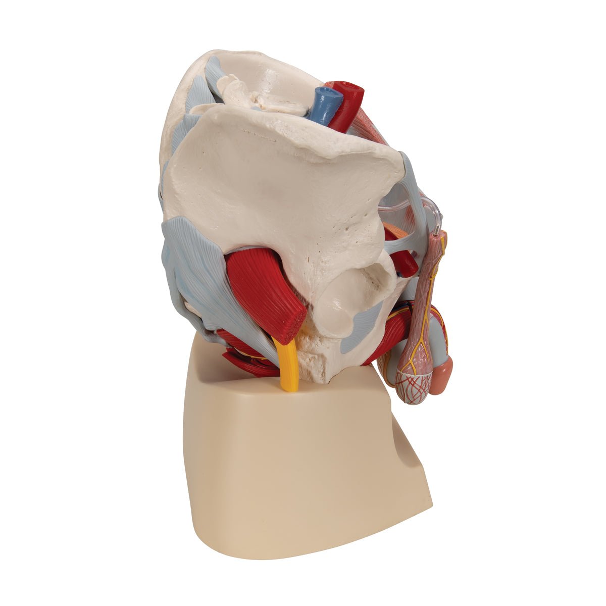
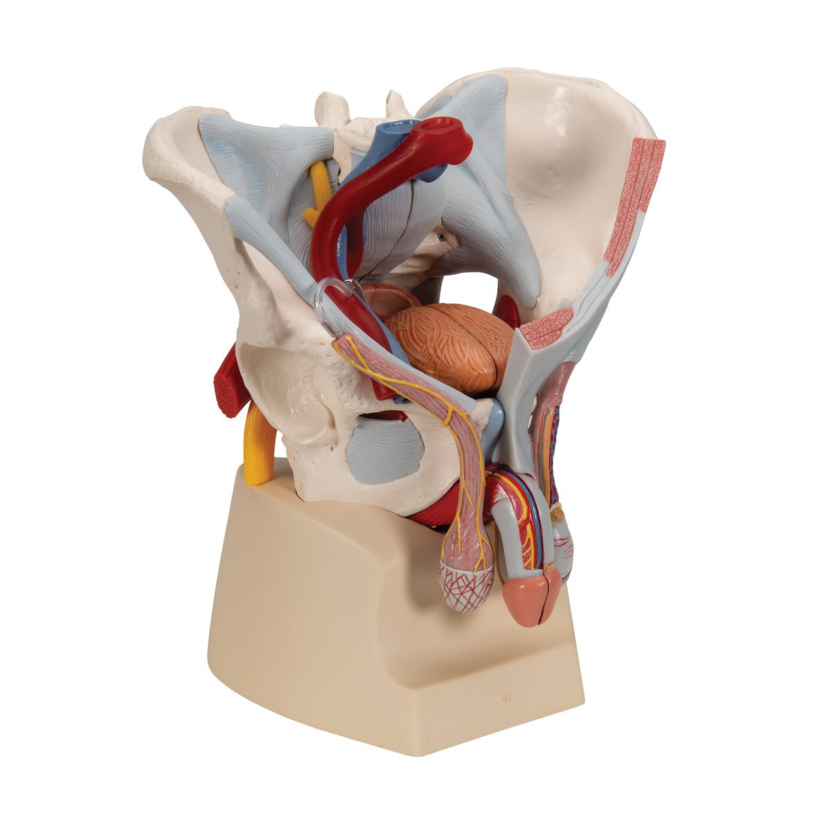
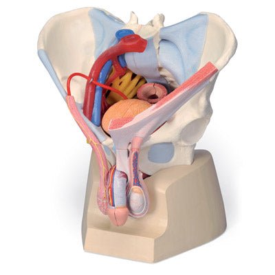
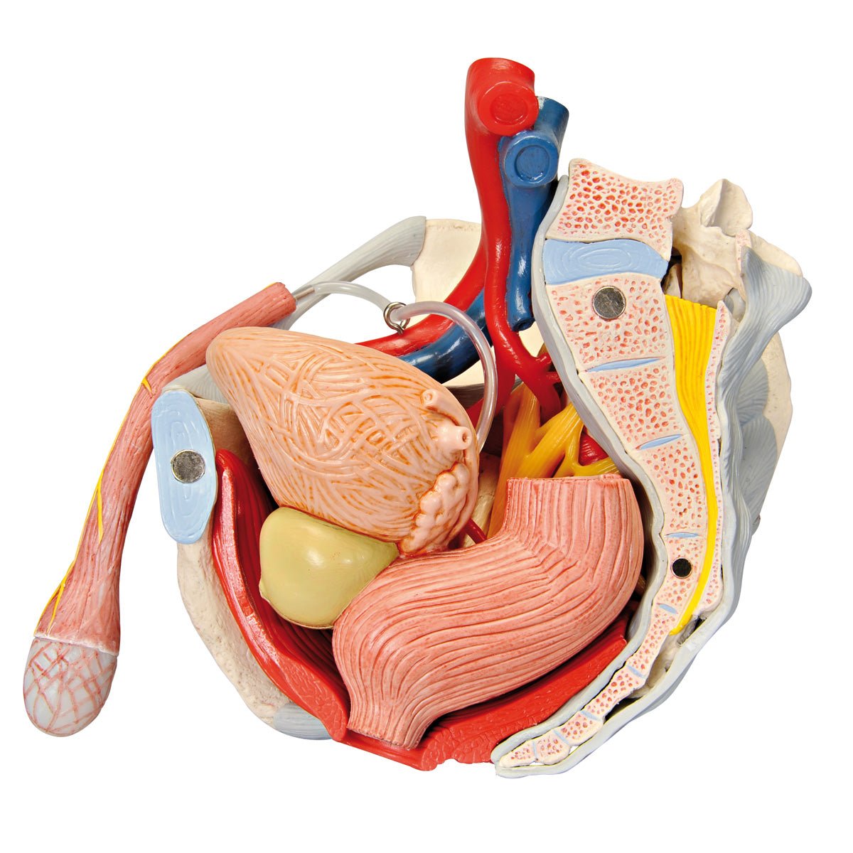
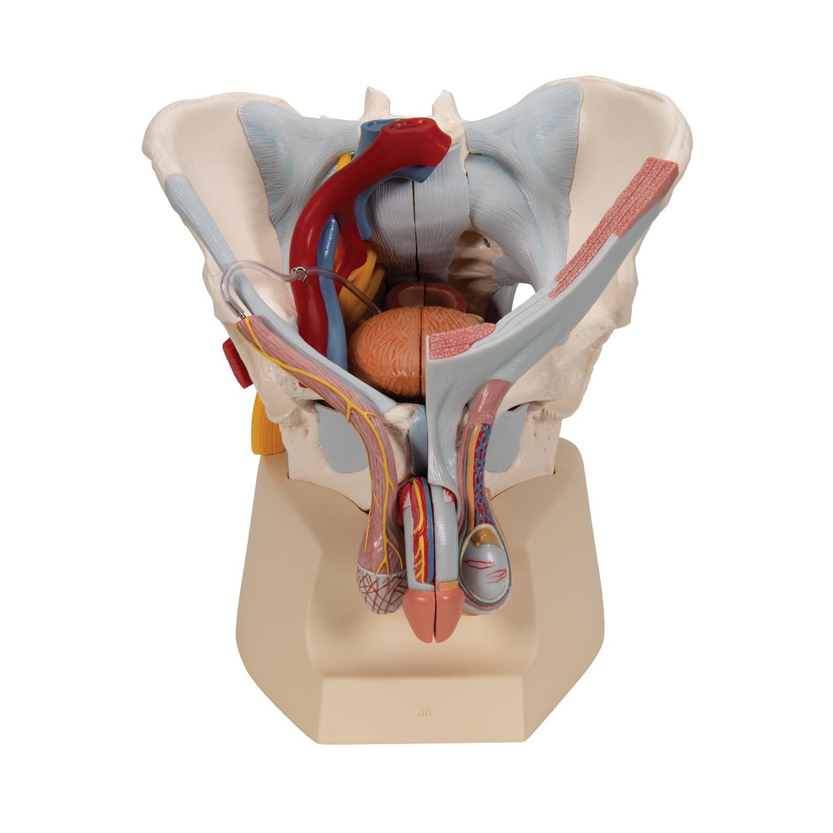
Pelvis with genitals, vessels and nerves, male, 7 parts

Item no.: 1013282
6.345,00 kr
Taxes included. Shipping calculated at checkout.

This product is shipped directly from the factory via UPS or DHL. When ordering, we will obtain the current delivery time. If the delivery time is more than 3-5 business days, we will contact you. You can also contact us for further information before ordering.

Description

This pelvis model shows the male pelvis with the following anatomical structures:


  • The pelvis (pelvic ring)
  • Pelvic floor (pelvic floor)
  • The internal genitalia
  • The spermatic cords
  • Some of the accessory gonads
  • The external genitalia
  • Ligaments, blood vessels and nerves in the pelvis
  • A bit of the abdominal muscles


The model is developed in natural adult size. It weighs approximately 3 kg and the dimensions are

21 x 28 x 31 cm. It can be separated into 7 parts (see the pictures on the left), which are held together using magnets and metal pins. The degree of detail on the bones is good, but there is no movement in the pelvic joints (e.g. the SI joints and the symphysis), although it e.g. can be split in half. The model is delivered on a stand.

Anatomically speaking

Anatomically speaking, the model shows many different tissues and organs, several of which can be seen in 3 dimensions because the model can be separated into 7 parts.


1) The pelvis consists of the 3 building blocks, which are the 2 hip bones and the sacrum. The model also shows the lower lumbar vertebra. In the bone tissue, the most important details can be seen, such as large nodules. Smaller details such as the linea glutaea are omitted. Overall, the model shows the following bones:


Os coxae (the 2 hip bones) of which both are composed
- Os ilium (iliac bone/hip bone)

- Os ischii (seat bone)

- Os pubis (pubic bone)


Us sacrum (sacrum) incl. os coccygis (coccyx)
5th lumbar vertebra (lumbar vertebra)


2) The pelvic floor (also called the pelvic floor or diaphragma pelvis in Latin) consists of the following muscles, which are clearly visible:


- M. levator ani (consisting of m. pubococcygeus and m. iliococcygeus)

- M. coccygeus


Other muscles are also seen such as the ischiocavernosus muscle, the bulbospongiosus muscle, the obturatorius internus muscle and the sphincter ani externus muscle. In addition, the relationships to the urinary bladder (vesica urinaria) and the rectum (rectum) are seen in detail.


3) The internal genitalia include the testicles (testes) and the epididymides, which are seen in relation to the scrotum and the spermatic cord (funicle). It is not possible to see the internal structures of the testicles (tubules, etc.). Both the contents of the spermatic cord and its barrier/leaves are clearly seen.


NB: In our assortment of pelvic models, we have 2 models that show the internal structures of the testicles: A large model with many details that can be viewed via THIS LINK and a miniature model that can be viewed via THIS LINK


4) The seminal ducts include the ductus deferens (the vas deferens), the ductus ejaculatorius and the urethra masculinum (the urethra), all of which are visible.


5) In relation to the accessory gonads, the prostate (bladder neck gland) and vesiculae seminales (seminal vesicles) are seen, but not gll. bulbourethrales and etc. urethral. The 2 first-mentioned glands are seen in cross-section, and therefore the excretory ducts and their connection are seen in detail.


6) The external genitalia include the penis and the scrotum, which are also seen. Opposite the testicles, the internal structure of the penis can be seen.


7) Ligaments, blood vessels and nerves are seen in detail. Ligaments are seen on both sides of the pelvic model, while blood vessels and nerves are mainly seen on the right side of the model.


Since the model i.a. can be divided in half, important details can be seen in a median section (including cauda equina). As for nerves, the sacral plexus, sciatic nerve (nervus ischiadicus) and other important nerves are also seen. As for blood vessels, the large artery and vein of the pelvis and parts of their branches are seen.


Many of the pelvic organs function as a channel or a reservoir (e.g. the rectum and the urinary bladder), which is why they are seen in a pedagogical way with an air-filled cavity (cavity).


8) Some of the abdominal muscles can also be seen on the model, so that the connection between the testes, the spermatic cords and the abdominal wall can be better seen.

Movement-wise

In terms of movement, the pelvis is not flexible. Although the various joints are visible (e.g. the SI joints and the symphysis), they are not movable.

Clinically speaking

Clinically speaking, the model can be used to understand diseases and disorders in the man's pelvis - for example in the genitals, the urinary bladder or the rectum. Examples are torsio testis, benign space-filling processes in the scrotum, urethral stricture, anal fissure, BPH (benign prostatic hyperplasia) and prostate cancer.


Furthermore, the model can be used to understand disorders and diseases such as cauda equina syndrome and deep vein thrombosis (DVT) in the pelvis.

CUSTOMER SERVICE

custom-made items

If the product is stated as a made-to-order item, it means that the product is of such a size or such a high quality level that the product is only available on order. Delivery time may vary, but the price will always remain the same! Contact us for more information if you wish to order.

Prices and payment methods
  • Prices are stated in DKK including VAT.
  • You can pay with;
    - EAN no.
    - MobilePay
    - Visa, Mastercard
Right of withdrawal & return
  • The right of withdrawal is 14 days from delivery.
  • Return postage is 60,- when purchasing a label from us (sent as a PDF file via email) or 0,- when returning in person to our address.
  • See our terms and conditions here
Delivery / pickup

If the item is listed as in stock , it is physically located at our address in Frederiksberg and can therefore be shipped or picked up. When ordering, choose what you want.

If you wish to pick up the item, this can often take place immediately after ordering, but must be agreed with us. If you wish to have it shipped , this will be done via Postnord and often the same day the order is received.

If the item is listed as ' shipped directly from the factory ', the delivery time is usually 2-5 business days, but depends on the factory's stock status. If you want more precise information, you can always contact us before ordering.

Facts about eAnatomy
  • Founded in 2004 as a sole proprietorship and converted to ApS in 2019
  • 100% owned by Christian Birksø who is also responsible for the daily operations.
  • Develops and markets both original products, designed and produced by eAnatomi, and distributes many international brands.
  • Sells to both private and commercial customers in Denmark and abroad.

Other products selected especially for you

Product categories

Raffle for free products

All recipients of our newsletter are entered into raffles for free products. Sign up today and be in the running!