


E
Eva Larsen Perfekt overblik

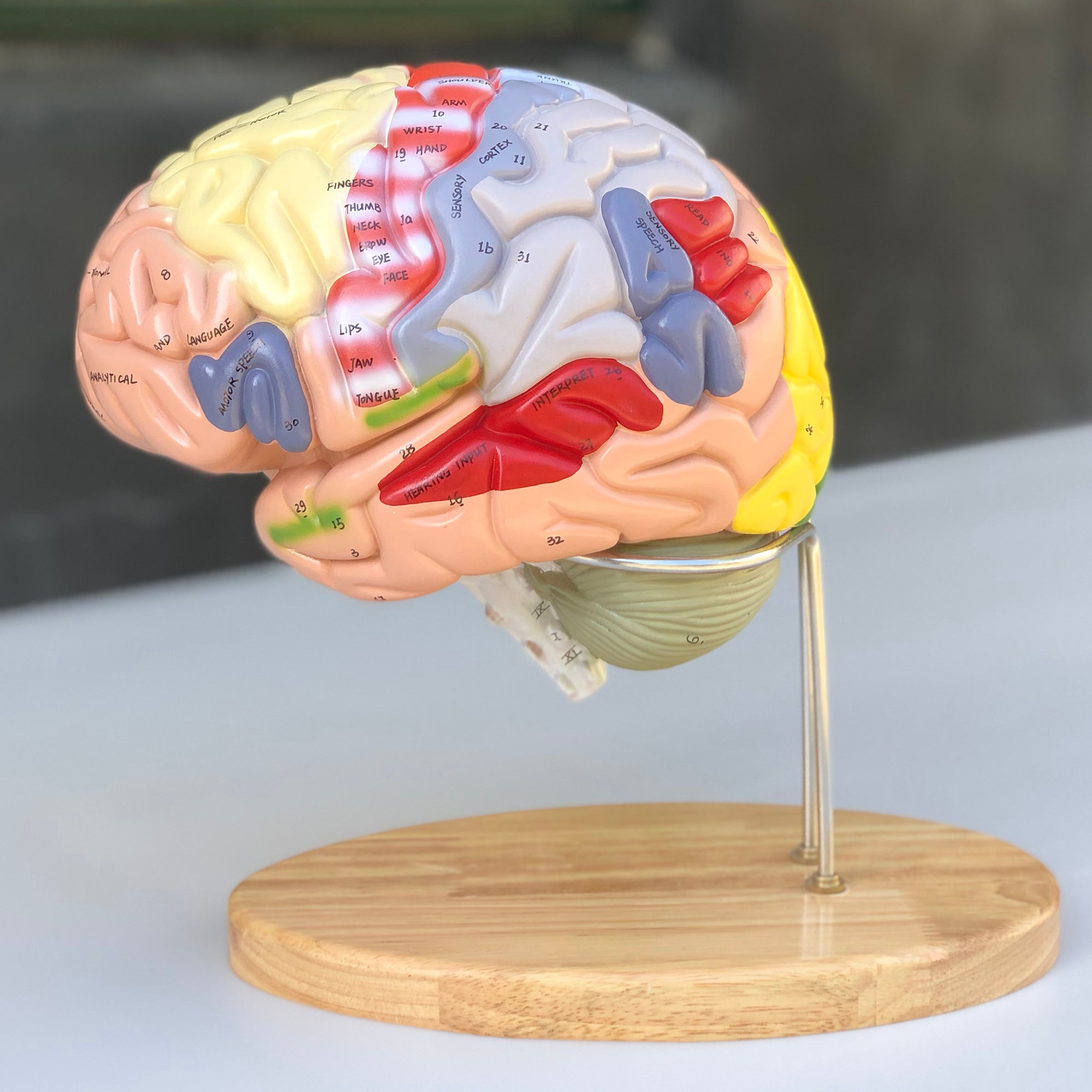
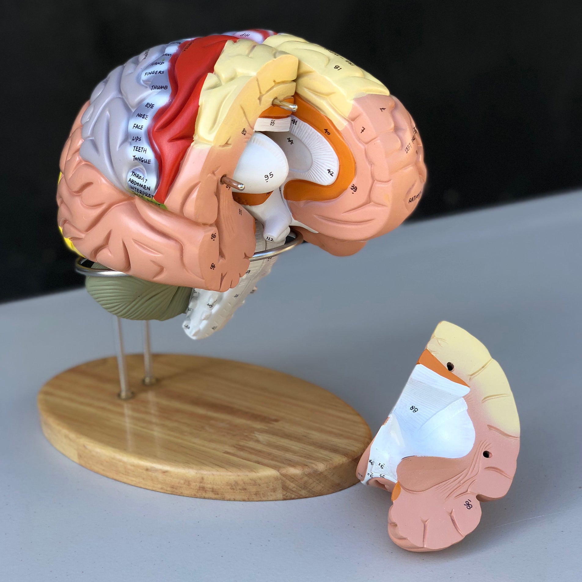
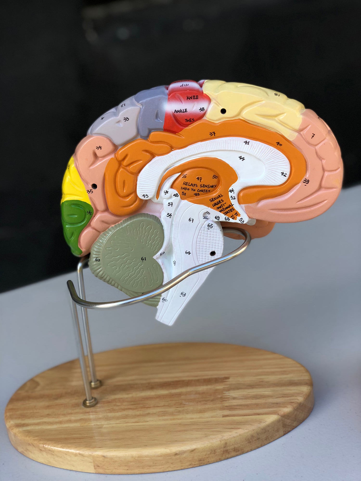
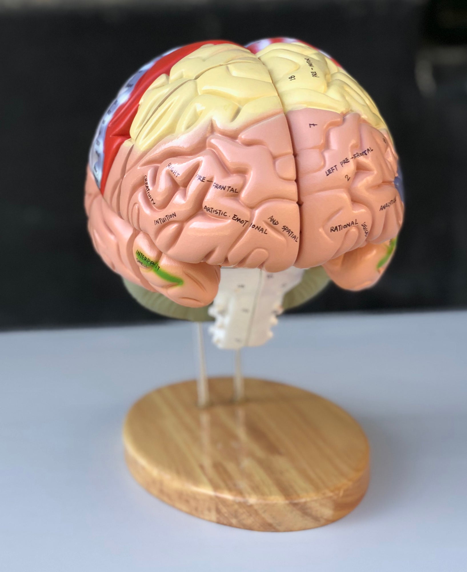
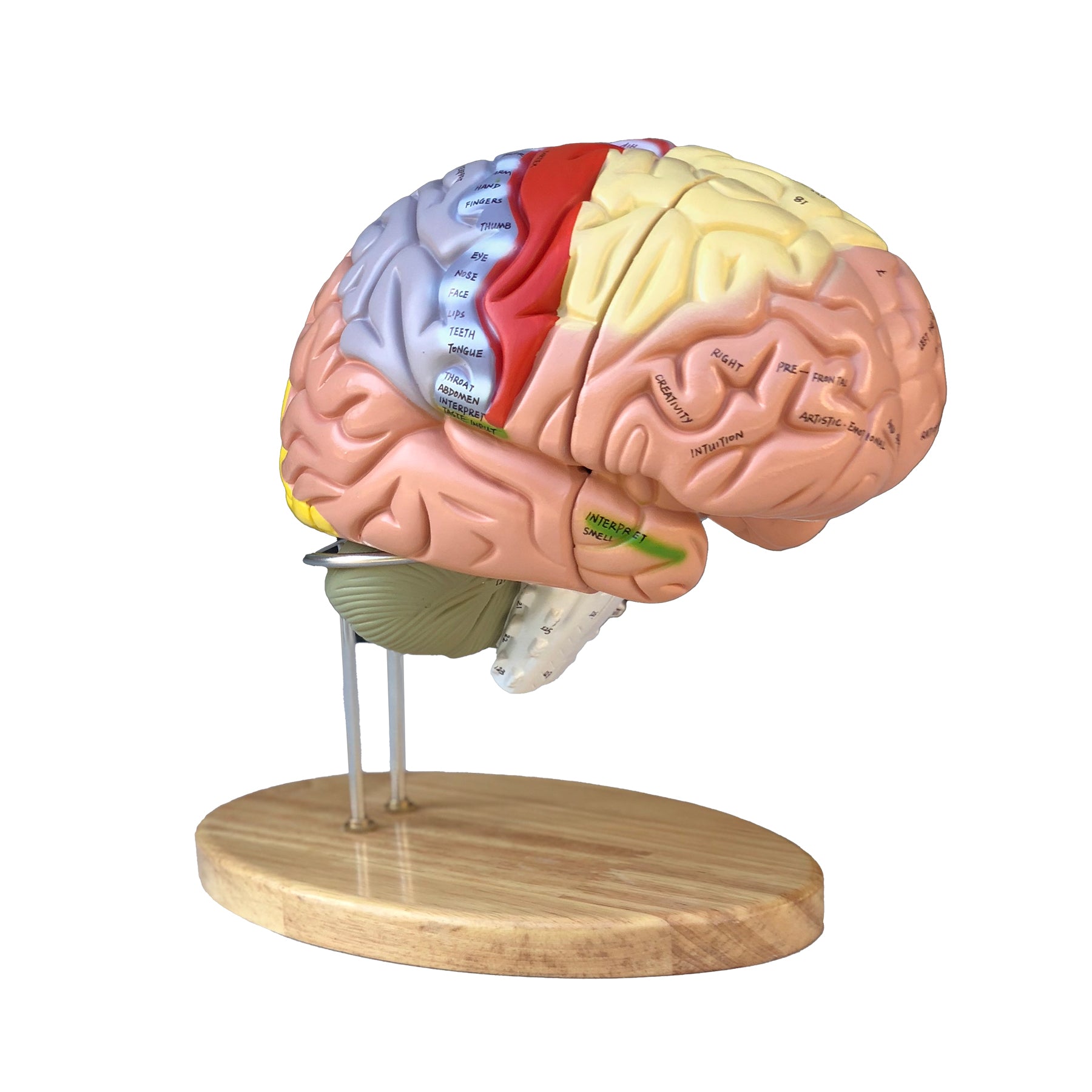
Brain, enlarged and educational anatomical model, 4 parts
Pickup available at Frederiksberg
Usually ready in 4 hours
If you are looking for an enlarged, inspiring and highly educational brain model that makes it as easy as possible to identify important areas in the brain, we highly recommend this one.
The model is cast in very hard and robust plastic. Unlike many of our other brain models, which are molded in hollow and flexible plastic, the material of this model can neither be pinched nor moved. Some would argue that this makes it less pleasant to touch and work with when it needs to be taken apart and studied. Others believe it is beneficial.
The model can be separated into 4 parts, which are held together via metal pins. Below you can read more about the anatomical details such as the colored areas and the limbic system. Compared to an adult, the model is approximately 1.5 times normal size. The dimensions are 22 x 18.5 x 19 cm and the weight is 1.9 kg. The model rests on a metal stand, which is mounted on a beautiful lacquered oak base with rounded edges (see pictures on the left). The model can be lifted off and separated very easily.
It should be emphasized that this brain model is the only one in our range that comes with both numbering of important anatomical structures as well as words written directly on special areas such as the primary motor area (see the images on the left and read more below).
NB: The numbering and naming are indicative. Therefore, be critical in your use, as figures for an anatomical structure may, for example, be located on the border of another structure.
CUSTOMER SERVICE
If the product is stated as a made-to-order item, it means that the product is of such a size or such a high quality level that the product is only available on order. Delivery time may vary, but the price will always remain the same! Contact us for more information if you wish to order.
If the item is listed as in stock , it is physically located at our address in Frederiksberg and can therefore be shipped or picked up. When ordering, choose what you want.
If you wish to pick up the item, this can often take place immediately after ordering, but must be agreed with us. If you wish to have it shipped , this will be done via Postnord and often the same day the order is received.
If the item is listed as ' shipped directly from the factory ', the delivery time is usually 2-5 business days, but depends on the factory's stock status. If you want more precise information, you can always contact us before ordering.
All recipients of our newsletter are entered into raffles for free products. Sign up today and be in the running!


