

Imagine coming face to face with a real human corpse. For many medical students, this moment is both thought-provoking and indispensable to their education.
This article examines how real cadavers are used in the education of doctors in Denmark in 2026, and why this method is still central, even though digital alternatives are gaining ground.
You will gain insight into the history behind cadaver dissection, the legal and ethical framework, the process from donation to teaching, and the latest teaching methods and alternatives.
Through the article, we create awareness about the learning value of the practice, challenge you with ethical dilemmas, and provide inspiration to understand future opportunities within anatomy education.
Historical background and development of cadaver dissection in medical education
Understanding how real cadavers are used in medical education requires a look at the fascinating history of cadaver dissection. This practice has shaped the development of medicine and left a deep mark on medical education, both in Denmark and internationally.
Development from ancient times to modern times
Already in antiquity, doctors began to examine the human body through dissection. In ancient Greece and Egypt, corpses were used to uncover the mysteries of anatomy, although the practice was often reserved for select circles. In the Middle Ages, dissection of corpses was banned in large parts of Europe, partly due to religious and cultural beliefs.
It was not until the 16th century that the use of cadavers for teaching was permitted again, especially after the groundbreaking work of Andreas Vesalius, who laid the foundation for modern anatomy. Vesalius insisted that knowledge of the body should be based on direct observation rather than ancient texts. This created a paradigm shift, with academic institutions gradually being allowed to perform cadaver dissections.
The transition from public dissections, where entire towns could witness the procedure, to closed, scientific sessions at universities marked an important development. In Denmark, cadaver dissection became a regular part of medical education from the 19th century, with the number of dissections increasing as universities grew. Statistics show that the number of dissections at Danish universities has increased significantly since the 20th century, in line with changes in legislation and an increased focus on practical learning.
The importance for medical science
Cadaver dissection has been crucial in answering the question: how are real cadavers used in medical education? Over the years, this practice has revolutionized the understanding of human anatomy and physiology. Many of the most important medical breakthroughs, such as the identification of the circulatory system and the discovery of the course of nerves, stem from meticulous dissections.
Compared to teaching on models or digital solutions, dissection of real cadavers provides unique insights. Students experience variations in anatomy that no model can reproduce, and gain a realistic feel for tissues and organs. This has a great impact on their practical skills.
Experienced teachers and students often emphasize that learning outcomes are greatest when theory and practice are combined at the dissection table. International and Danish studies document that the use of real cadavers strengthens both understanding and respect for the body. For students who want to delve deeper into the subject, there are specialized resources, such as For students in medicine , where you can find additional information about practice and materials for medical education.
In summary, cadaver dissection has helped shape both medical knowledge and the education of future doctors. By examining how real cadavers are used in medical education, it becomes clear that this practice continues to play a central role in equipping the healthcare professionals of the future.
Legal and ethical framework for the use of real corpses
Understanding how real cadavers are used in the education of doctors requires insight into both the legal and ethical frameworks that govern practice in Denmark. These frameworks ensure that the process of donation, storage and use of cadavers is carried out with respect and responsibility. The following is a review of both the legislation and the key ethical considerations that characterise the societal debate.
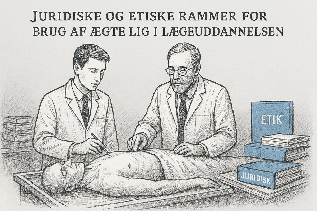
Legislation and guidelines in Denmark
In Denmark, the use of real cadavers in medical education is strictly regulated by law. The Health Act, especially Section 187, sets out the basic requirements for how real cadavers are used in the education of doctors. The central principle is that donation can only take place on the basis of written and informed consent from the donor himself. Relatives must be informed, but cannot oppose the donor's wishes, unless special circumstances apply.
Universities and hospitals are responsible for the proper receipt, storage and use of donated bodies. Storage is carried out under strict hygienic and ethical guidelines, where anonymity and dignity are prioritized, among other things. Authorities supervise, and there are detailed procedures for the transport and preservation of bodies.
Statistics show that the number of donated cadavers in Denmark has been stable in recent years, with approximately 150-200 donations annually. This covers the need for Danish universities' anatomy teaching, but there are occasional periods of shortage. To ensure correct practice, the universities follow the current national guidelines, which are continuously updated.
Here you can read more about the legal provisions in Section XIII of the Danish Health Act on postmortem examinations and autopsy , where requirements for consent, storage and use are described.
The legislation also takes into account how real corpses are used in the training of doctors in relation to research and further education. Here, separate permits and documentation are required so that the donor's will is always respected.
Ethical considerations and societal debate
The ethical framework for how to use real bodies in the education of doctors is at least as important as the legal one. Respect for the donor's will is fundamental. Many donors consciously choose to contribute to medical education, and this is seen as an altruistic act. The role of relatives is primarily to be informed and support the process, but occasionally difficult situations arise where emotions and dignity come into play.
The debate about dignity and anonymity is a hot topic in the public sphere. Corpses are treated with the utmost respect, and there are clear rules for how they are handled in education. Students learn early on to show respect and understanding for the importance of their work. This is part of the education and professional ethics that accompany medical education.
International differences also shape the debate. In some countries, it is forbidden to use real corpses, while others have more lenient rules. Denmark balances between ensuring high quality in education and protecting the rights of donors and relatives.
Ethical dilemmas arise especially when there is disagreement about the interpretation of the donor's wishes, or when the media and society question the practice. Educators and ethicists contribute to the debate on an ongoing basis, and there is constant work to improve procedures and communication.
Overall, the ethical and legal framework is about ensuring that the use of real cadavers in medical education is done with respect, responsibility, and societal acceptance. It is this balance that makes the practice possible and meaningful for both students and society.
The process: From donation to dissection
Understanding the process behind how real cadavers are used in medical education requires an insight into each individual step. The entire process begins long before the actual teaching, and each step is influenced by both ethical, legal and practical considerations. Below, we review the three central phases that ensure that real cadavers are used responsibly and with dignity in medical school.
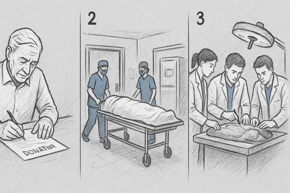
Step 1: Donor decision and consent
The first step in how to use real cadavers in the education of doctors is the donor's personal choice to donate his body to science. This decision is often made well in advance and involves a thorough information process. The donor must fill out and sign a legally binding consent form, which is registered with the relevant authorities. It is important that the donor's wish is clearly formulated and documented so that no doubts arise later in the process.
Family and relatives are typically informed of the decision, as they can be involved if questions arise after the death. Although the decision is personal, many choose to discuss the wish openly to ensure respect for all parties. Statistics from recent years show that around 200 people annually in Denmark choose to donate their body for medical education. However, the number can vary from year to year, depending on societal debate and awareness campaigns.
The donation process follows strict legal frameworks and national guidelines. For those interested, you can read more about National recommendations for donation after circulatory death , where the process is described in detail. This ensures that how real cadavers are used in the education of doctors always takes place under controlled and ethically sound conditions.
Step 2: Receipt and preparation of the body
When a death has occurred and it is confirmed that the deceased wanted to donate their body, the next phase of how real cadavers are used in the education of doctors is initiated. The body is transported safely to a university hospital or an anatomical institute. There, it is received by specially trained personnel who handle the body with great respect.
First, the body is registered and a thorough identification is made. Then, preservation begins, which typically involves the use of formalin or other chemical agents to ensure that tissues and organs are preserved for educational use. Alternatively, freezing techniques can be used if certain anatomical structures are desired to be preserved more naturally. These processes are essential for the body to be used safely and effectively for a longer period of time.
The safety of both staff and students is given high priority. Requirements are made for ventilation, protective equipment and correct handling of chemicals. The process is carefully documented so that everyone can follow how real corpses are used in the training of doctors in practice. Experience from Danish universities shows that an average body can be used in teaching for up to a year, depending on the preservation method and teaching needs.
Step 3: The actual dissection in teaching
The final phase of how to use real cadavers in medical education takes place in the classroom itself. Here, the body is typically divided into regions so that multiple teams of students can work on different areas simultaneously. Each group is given responsibility for a specific part – for example, the thorax, abdomen or extremities – which allows for specialized learning.
The dissection is conducted under the guidance of experienced instructors. Students gain hands-on experience with anatomy, tissue structure and organ placement, which cannot be achieved through digital models alone. The practical work is supplemented by theoretical lectures, where observations from the dissection are discussed. The teaching is carefully planned, and a logbook is kept of each group's progress.
For many students, it is an emotional challenge the first time they are faced with a real human corpse. Respect and ethics are emphasized in the teaching, and students are encouraged to reflect. Statistics from Danish medical studies show that an average of 40-60 hours are spent per student on dissection during basic education. This intensive course contributes to how real corpses are used in the education of doctors providing a unique and irreplaceable learning outcome.
Teaching methods and learning outcomes
Teaching methods play a crucial role in how real cadavers are used in medical education. The combination of practical experience and theoretical knowledge ensures that future doctors gain a deep understanding of the anatomy of the body. It is precisely in this area of tension that real cadavers continue to have a central place, despite new technological alternatives.
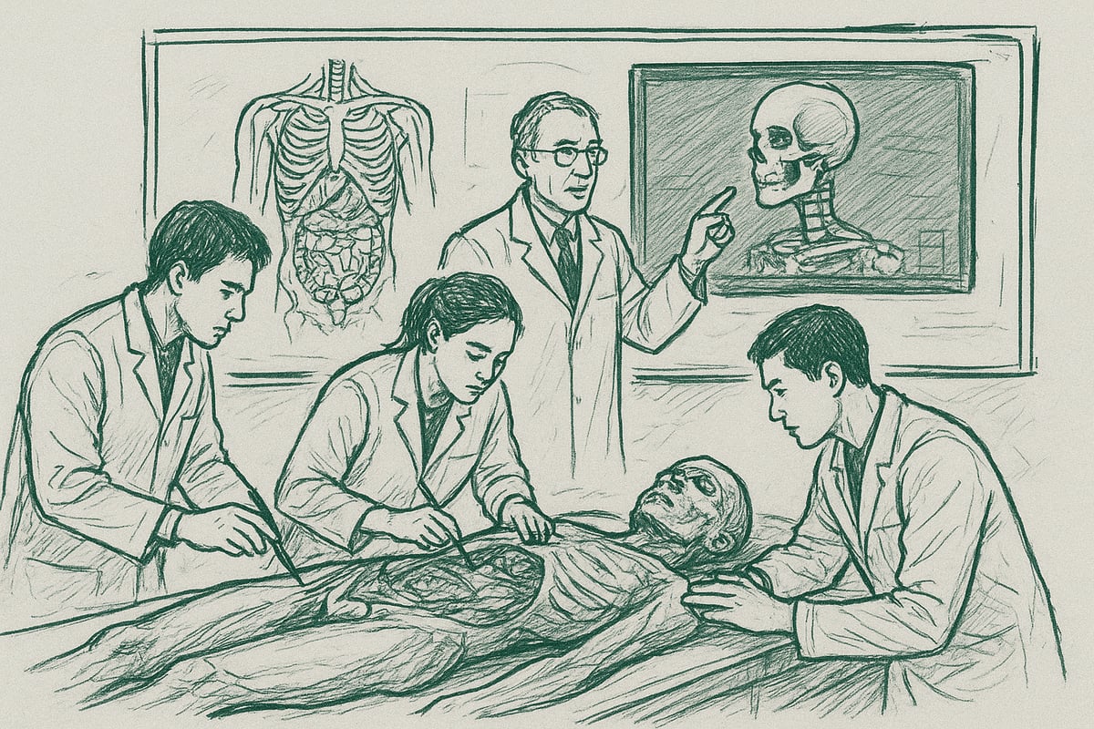
Practical techniques and pedagogy
When looking at how real cadavers are used in medical education, hands-on dissection is still the most widely used method. Students are given the opportunity to examine the structures of the body directly, providing a tactile and three-dimensional understanding that cannot be achieved through digital models alone.
In addition to the dissection, demonstrations are also used, where the teacher shows specific anatomical areas. This method is used especially to highlight complex relationships that may be difficult to uncover individually. Theory and practice are often integrated, so that students connect book knowledge to concrete observations on the body.
Interdisciplinary teams, where anatomy, surgery and pathology work together, enhance understanding of body function and disease. Several universities are experimenting with innovative teaching programs where students actively participate in both planning and performing dissections. Perspectives from both educators and students show that this varied approach promotes engagement and learning.
An important resource is access to anatomy teaching materials that complement dissection with digital tools and models. This ensures that teaching can be adapted to different learning styles and needs, and makes it easier to prepare for clinical situations.
Documented effect on learning and skills
Research shows that the use of real cadavers in medical education has a significant impact on both theoretical and practical skills. Studies often compare learning outcomes from cadaver dissection with digital alternatives. The results indicate that students who work with real cadavers generally gain a deeper understanding of the anatomy and variations of the body.
A review of exam results from Danish universities shows that students with practical experience in dissection perform better in both theoretical exams and clinical skills tests. At the same time, many report that the experience of respect and ethics in teaching is strengthened when working with real human bodies. This aspect is central to understanding how real cadavers are used in medical education and why this tradition continues to be prioritized.
However, there are challenges. Some students experience emotional strain, especially the first time they work with corpses. The resource requirements for storage, security and teaching are significant, which can challenge the capacity of universities. Despite these challenges, educators assess that the learning benefits outweigh the costs, especially when it comes to understanding human complexity.
In conclusion, the experiences of both students and educators illustrate how using real cadavers in the education of doctors remains an indispensable part of anatomy teaching, even in an era of rapid technological development.
Alternatives to using real corpses: Advantages and limitations
As technological advances progress, many are asking the question: how are real cadavers used in medical education when alternatives are gaining ground? Modern teaching environments increasingly combine classical methods with digital solutions and artificial models. This development opens up new opportunities for learning, but also requires evaluation of what the different methods can actually offer.
Artificial models and digital solutions
Artificial models and digital solutions are playing a larger role in how real cadavers are used in the education of doctors at Danish universities. 3D-printed organs, plastinates, virtual reality (VR) and augmented reality (AR) allow students to explore anatomy without physical contact with real bodies.
These alternatives offer several advantages:
- Safety: No risk of infection or chemicals.
- Availability: Can be used repeatedly and shared across teams.
- Ethics: No ethical dilemmas associated with the use of human remains.
However, the alternatives also have certain limitations:
- Lack of realism: Artificial materials cannot fully mimic the variations of the human body.
- Tactile feedback: The feel of tissues, organs, and structures cannot be recreated digitally.
- Variation: Real bodies show natural variation that models cannot always illustrate.
A concrete example is 3D dissection models as an alternative , which are becoming more popular in anatomy teaching. Statistics from Danish universities show that up to 30 percent of teaching time can now be spent with digital or artificial alternatives, especially in the early semesters.
Comparison with real corpses
When considering how to use real cadavers in medical education, many educators point out that certain skills are best trained through the actual dissection. Handling real tissue, understanding individual anatomical variations, and building respect for the human body are difficult to transfer to digital platforms.
Research shows that students often prefer a combination of methods. Models and VR help visualize complex structures, but many find that hands-on work with real cadavers provides a deeper understanding. Clinical skills, such as precise dissection or surgical technique, are best strengthened through the authentic experience.
Perspectives from both educators and students emphasize that digital alternatives are a valuable supplement, but cannot stand alone. Optimal teaching occurs when the different tools are combined, so that they each contribute to answering the question: how can real cadavers be best used in the education of doctors?
| Method | Benefits | Limitations |
|---|---|---|
| Real corpse | Realism, variation, tactile feedback | Resource-intensive, ethical challenges |
| 3D/VR/Models | Security, accessibility, ethics | Lack of realism, limited emotion and variety |
In summary, the development shows that the balance between new technologies and the use of real cadavers must be adjusted to both learning objectives and ethical frameworks. This requires ongoing evaluation to ensure that the doctors of the future are equipped with the necessary competencies.
The Future of Anatomy Teaching: Trends and Visions for 2026 and Beyond
How will real cadavers be used in the education of doctors in the future? This question is central to the development of anatomy education in Denmark. As technology and social conditions change, the field faces new opportunities and challenges. The future offers a balance between tradition and innovation, where both learning and ethics weigh heavily.
Development in technology and teaching methods
In 2026, it is clear that how to use real cadavers in medical education is no longer just about classic dissection. Artificial intelligence, virtual reality and 3D models have been integrated into teaching. Many universities are now combining traditional methods with digital solutions. This allows students to alternate between hands-on experience and advanced simulation.
International trends indicate that hybrid education will become the norm. Both Danish and foreign universities are experimenting with projects where students first practice virtually and then work with real tissue. This strengthens the understanding of complex anatomy and allows for individual preparation.
Examples from Denmark show that collaboration between disciplines such as anatomy, surgery and technology is growing. New teaching materials explaining how to use real cadavers in medical education are readily available to students. For those who want more knowledge about materials and an introduction to the field, resources are available at For the new medical student .
This development means that students have more learning paths. Technology complements, but cannot completely replace, the experience of working with real bodies. It is precisely the combination that creates deep understanding.
Ethics, resources and sustainability
When asking how real cadavers will be used in the education of doctors in the future, ethics and sustainability play an increasingly important role. The extent of body donation is expected to change, partly due to increased public awareness and new alternatives. Environmental considerations also become important, as the preservation and transport of cadavers have a climate footprint.
Sustainable alternatives such as plastinates and digital solutions are gaining ground, but they cannot yet fully replace the learning value that real corpses offer. Experts emphasize that the balance between respect for the donor, quality of learning and the environment must be prioritized.
The societal debate about how real cadavers are used in medical education involves both ethical dilemmas and practical questions. Should the use of real cadavers continue, or should we focus more on digital alternatives? Perspectives from students and educators show that dignity and consent remain central themes. For a deeper insight into the ethical considerations, we recommend Ethical aspects of research in health data , where principles for the use of biological material are discussed.
The future requires ongoing dialogue between professionals, society and authorities. Only in this way can we ensure that the anatomy education of the future remains both innovative and responsible.
When we look at how real cadavers are used in medical education, it becomes clear how important both historical traditions, today's ethical frameworks and technological solutions are for the education of the future. If you are curious to explore how anatomical models, posters and innovative learning tools can complement or perhaps even change the way we understand anatomy, you can find inspiration and knowledge at eAnatomi ApS. Here you will have access to a wide range of resources that make complex topics accessible to everyone, regardless of whether you are a student or a professional.
Read more here
0 comments