
How do you really learn about the anatomy of the heart? A heart model allows you to see, touch, and understand the complex structures that keep us alive. It's key to both health and effective learning.
This guide shows you how the latest heart model in 2025 can make anatomy tangible and revolutionize your approach to learning. We review the model's construction, its use in education and the healthcare sector, choosing the right model, practical use and future trends.
Are you curious about how a heart model can make your learning easier and more effective? Read on and discover how you can get more out of your understanding of the heart.
What is a heart model?
Understanding how the heart works requires more than just pictures in a textbook. A heart model brings anatomy to life and allows you to experience the heart's complex structures in a tangible way. As teaching methods evolve, heart models have become indispensable in both education and healthcare.
Definition and purpose
A heart model is a physical or digital representation of the anatomy of the heart, designed to provide a detailed and realistic understanding of the structure and function of the heart. Unlike general anatomical models, a heart model specifically shows the chambers, valves, vessels and often also pathological conditions of the heart. This precision makes the heart model a key tool for educators, students and healthcare professionals.
The purpose of a heart model is to visualize the structure of the heart, the path of blood and the mechanisms that control the heart's function. Historically, heart models have evolved from simple plastic versions to advanced 3D versions that can be disassembled and show even the smallest details of the heart's anatomy. Today, the focus is on interactivity and realism, making heart models an effective supplement to both traditional and digital forms of learning.
The use of heart models is wide-ranging. In education, they provide the opportunity to demonstrate blood flow, explain disease processes and train clinical skills. In research, they are used to test new techniques, while in patient communication they help explain diagnoses and treatments in an easily understandable way. According to Heart Models for Education, there is an increasing demand for anatomical models, especially because they promote learning and make complex relationships more manageable.
Compared to digital simulations, a physical heart model provides a unique sensory and in-depth learning experience. Being able to hold the model in your hand, take it apart, and look inside the heart significantly increases understanding. Research shows that tangible learning improves both the retention of knowledge and the ability to apply it in practice. That is why many educators still choose physical models as a supplement to digital solutions.
Types of heart models
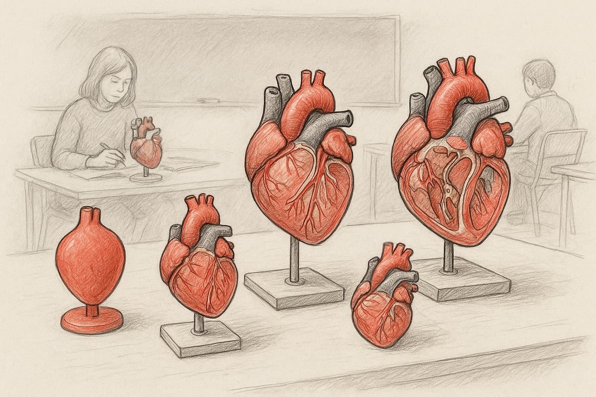
The range of heart models ranges from simple to highly sophisticated. The classic life-size heart model is popular in basic education, where it provides a realistic picture of the heart's proportions. Enlarged models are often used for detailed teaching, while reduced models can be practical for transport or patient guidance.
There are models that focus on specific aspects such as the impulse system, heart valves or pathological conditions such as atherosclerosis and hypertrophy. Special models include fetal hearts, hearts with pericardium or models illustrating bypass surgeries. Some models are two-part and provide access to the internal structures, while others consist of up to 12 parts for maximum detail.
The choice of materials varies from robust plastic to soft silicone and advanced 3D printing. Plastic models are durable and suitable for everyday use, while silicone provides a more realistic feel. 3D printed models allow for tailor-made solutions for specific teaching or research needs.
The use of different models often depends on the level of education. In higher education and clinical training, there is a greater demand for models with a high level of detail and the possibility of interactivity. At the undergraduate level, simple models are often used to provide an overview without overwhelming students.
Each type of heart model has its advantages and disadvantages. A simple model is easy to use and understand, but has limited detail. An advanced model provides deeper insight, but requires more time and explanation. The choice therefore depends on the purpose, user group and learning goals.
Structure and function of the heart model
Understanding the complex anatomy of the heart requires more than just pictures and text. A heart model allows you to physically experience the heart's structures, which enhances both learning and understanding. The right heart model can bring theory to life and make even the most difficult relationships clear.
Anatomical details and precision
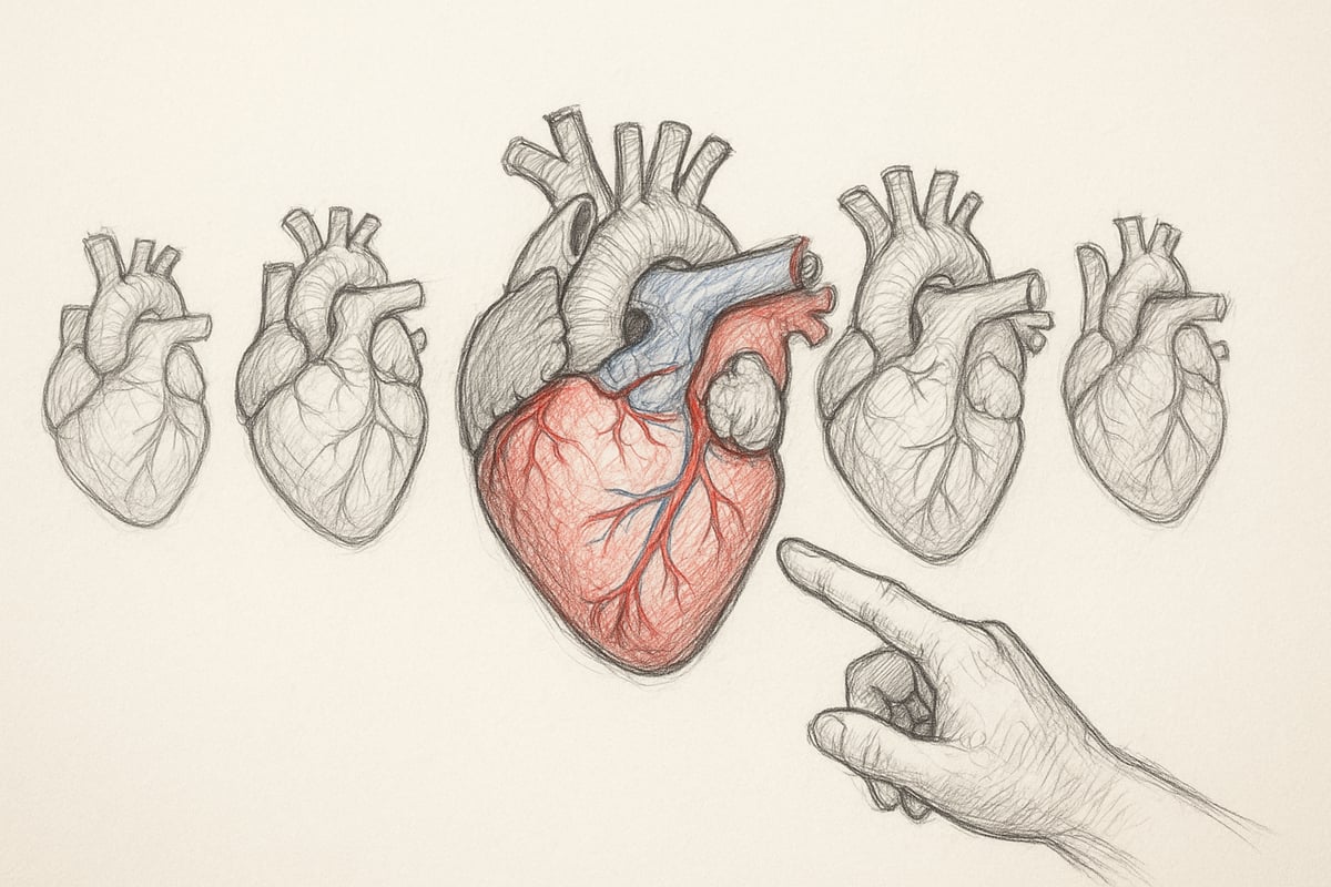
A heart model is constructed with a focus on reproducing the main structures of the heart: atria, ventricles, valves, vessels and pericardium. Precise anatomical details, such as color coding and removable parts, make it possible to follow the path of blood through the heart.
When a heart model can be disassembled, the user can examine the inside and see how the valves and chambers work together. For example, 2-part models show the essential internal structures, while 12-part models allow access to even the smallest anatomical details.
| Model type | Number of parts | Focus area | Target group |
|---|---|---|---|
| 2-piece | 2 | Basic anatomy | Student |
| 12-piece | 12 | Detailed structure | Specialists |
| Pathology model | 4-6 | Disease course | Patient education. |
| Fetal heart | 2-3 | Development phases | Health students |
Accuracy in a heart model is crucial, especially in medical education and simulation training. The more detail the model has, the better the user understands both normal conditions and pathological changes, such as atherosclerosis or hypertrophy. A detailed heart model also helps to visualize relationships that may be difficult to explain with images alone.
Interactivity and usability
An interactive heart model allows for the individual parts to be examined and assembled, making teaching more engaging. Models that can be disassembled are often used to demonstrate surgical procedures or disease progression.
The use of heart models in patient education and simulation training ensures that both students and instructors can work with concrete examples. This not only increases understanding, but also learning, as studies show that physical models promote engagement in teaching.
Compared to digital alternatives, a physical heart model provides a unique, tactile learning experience. However, when combined with digital solutions, it can provide an even broader learning outcome. Statistics indicate that the use of physical models in education has been increasing, especially in healthcare education, where a heart model is often considered indispensable.
eAnatomi ApS: Experts in anatomical heart models
eAnatomi ApS is a specialist in the development and distribution of anatomical models and posters, including heart models, for both educational institutions and the healthcare sector. The company has a close collaboration with healthcare specialists, which ensures high professional precision and quality.

The selection ranges from classic 2-part heart models to advanced models illustrating the impulse system and various pathological conditions. You will find life-size models, enlarged versions and special models for specific needs, such as patient communication and surgical training.
eAnatomi ApS delivers globally and offers solutions to both private and professional users. Their focus is to create learning through tangible products that strengthen the understanding of complex anatomical conditions. If you want to know more about the company's background and expertise, you can read more at About eAnatomi ApS .
The benefits for educational institutions and professional users are clear: access to quality models that support both theory and practice, as well as a solid partner with extensive experience in the field.
Choosing the Right Heart Model: Step-by-Step Guide
Choosing the right heart model can be crucial for both learning outcomes and practical applicability. A structured approach ensures that the model matches your needs, whether you work in education, healthcare or research. Here is a step-by-step guide to help you make the best choice.
1. Identify the purpose of the model
The first step is to define what you want to achieve with your heart model. Will it be used for teaching, patient education, or perhaps research? If you are teaching students, a model with visible diseases or surgical procedures can make a big difference. For example , heart models for students can help reinforce understanding of complex topics through visual and tactile learning.
Also consider whether you need a model that illustrates specific conditions such as atherosclerosis or heart valve disease. The right choice depends on who the target audience is and what anatomical or clinical aspects you want to highlight.
2. Choose the appropriate size and level of detail
Once the purpose is determined, you should consider the size and level of detail of your heart model. Life-size models are well suited for individual teaching or patient dialogue, while enlarged models can enhance understanding in larger groups. Reduced models are convenient for transportation or overview.
The level of detail varies from simple models to advanced versions with many removable parts. Consider whether you need to be able to show the path of blood through the heart, or whether the focus is primarily on superficial structures. For in-depth courses, detailed models are often preferable.
3. Consider materials and durability
The choice of material has a great impact on both ease of use and longevity. The most common materials for a heart model are plastic, silicone and 3D printing. Each type has advantages and disadvantages, which can be summarized in the table below:
| Material | Advantages | Disadvantages |
|---|---|---|
| Plastic | Affordable, lightweight | Less realistic |
| Silicone | Realistic, flexible | More expensive, requires care |
| 3D printing | Tailored, precise | Can be fragile |
If the model will be used frequently or cleaned frequently, it is important to choose a material that can withstand daily use. Also consider whether the model will be able to withstand disinfection.
4. Interactivity and usability
An interactive heart model that can be disassembled and reassembled provides significantly better learning opportunities. The ability to show internal structures and simulate disease processes or surgical procedures increases understanding.
Consider how easy the model is to handle for both teachers and students. Some models come with a stand or storage case, making them easy to transport and protect. Ease of use is especially important if the model is to be used by several different user groups.
5. Price and budget considerations
The price of a heart model varies considerably, depending on size, level of detail and choice of materials. For educational institutions, it may be advantageous to obtain quotes and investigate opportunities for discounts on larger purchases.
Always compare several models and assess whether a simple model will meet your needs or whether it is worth investing in a more advanced version. Remember to include any costs for maintenance and spare parts in your overall budget.
6. Certification and quality assurance
Quality and safety should always be a top priority when choosing a heart model. Check to see if the model is CE marked and meets applicable medical device standards. Certified models ensure that the anatomy is accurately reproduced, which is essential for both teaching and clinical use.
Collaboration with healthcare specialists during the development of a heart model is often a seal of quality. Choose a supplier who can document high professional precision and durability. This way, you will get a heart model that is both safe and provides maximum learning benefits.
Heart models in education and healthcare
Understanding the complex anatomy of the heart requires more than just theory. A heart model can make the difference between abstract knowledge and real insight. This section highlights how heart models are used in education, patient communication, and research, and why they are indispensable tools in the healthcare sector.
Use in education
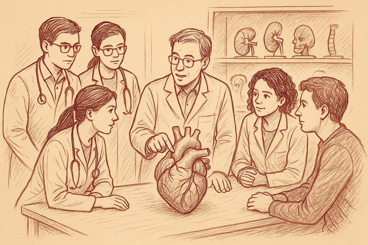
Heart models are today a central tool in both basic and advanced health education. Medical students, nurses and physiotherapists are given the opportunity to examine the structures of the heart in detail through practical exercises. By using a heart model, they can simulate blood flow, identify valves and vessels and understand disease development.
A statistical trend shows that the use of heart models in anatomy teaching has increased significantly in recent years. This is partly because tangible learning increases both understanding and retention of knowledge. For example, many students find that they can more easily remember the structure of the heart when they can physically take the parts apart.
List of benefits of using a heart model in teaching:
- Provides deeper anatomical understanding
- Promotes active learning and engagement
- Supports collaboration between students
A heart model is therefore not just a supplement, but a necessity for effective and modern teaching.
Patient communication and understanding
When healthcare professionals need to explain diagnoses and treatments, a heart model becomes an invaluable tool. The model makes it possible to visualize, for example, a bypass operation or atherosclerosis, so the patient can see where the problem occurs. This not only strengthens understanding, but also increases the patient's motivation to follow the treatment plan.
Studies document that patients who are presented with diseases via a heart model are better able to understand their situation and make informed choices. According to the Danish Health Authority's report on cardiac follow-up 2024, patients experience a higher degree of security when information is provided visually and tangibly.
Examples of use:
- Explanation of heart valve diseases and surgeries
- Visual demonstration of risk factors for atherosclerosis
- Preparing the patient for surgical procedures
A heart model acts as a bridge between healthcare professionals and patients, promoting dialogue and compliance.
Research and innovation
Cardiology research greatly benefits from heart models in the development of new treatments and medical devices. By testing surgical techniques on models, researchers and doctors can safely test procedures before clinical use. A heart model allows for the simulation of different anatomical variations and disease patterns, which enhances the innovation process.
This practice minimizes the risk of errors and contributes to more effective and safe treatment. In addition, a heart model can be used to illustrate complex conditions in collaboration between different healthcare teams.
In summary, heart models are indispensable in both daily work and in the development of future cardiac treatments.
Practical use and maintenance of heart models
A heart model is an indispensable resource in both education and healthcare. To ensure optimal lifespan and function, proper use and maintenance are essential. This is not only about storing the model properly, but also about daily handling, cleaning, and understanding the durability of the materials. By following a few simple guidelines, you can significantly extend the lifespan of your heart model and ensure that it remains an effective learning tool.
Daily use and storage
To maintain the quality of your heart model, handle it with both hands and avoid pulling on small parts. It is recommended to place the model on a stable, dry surface when not in use. Store the heart model away from direct sunlight and moisture, as these factors can degrade the material over time.
Cleaning should be done with a soft, slightly damp cloth and a mild detergent. Avoid aggressive chemicals as they may damage the surface. After cleaning, the model should be allowed to air dry completely before packing away. For institutions, it is advantageous to have regular routines for daily inspection and cleaning.
When stored in classrooms, the heart model should be placed in a dust-proof box or cabinet. In clinics, it is important to ensure that the model is always easily accessible, but at the same time protected from accidental contact or loss. Proper storage extends the life of the model and ensures that it is always ready for use.
Troubleshooting and repair
Even the most robust heart model can show signs of wear over time. Typical problems include loose parts, discoloration, or minor cracks. If a part comes loose, you should immediately check whether it can be fixed without the use of glue, as some materials can be damaged by strong adhesives.
A heart model with discoloration can often be cleaned with a special plastic or silicone cleaner. Avoid hard brushes, which can scratch the surface. For minor damage, small repairs can be made with materials that match the model's original composition.
If the heart model becomes extensively damaged, you should consider replacing it. Investing in a quality heart model pays off as it lasts longer and requires fewer repairs. Keep an eye on any changes in functionality so you can intervene quickly to prevent further damage. For institutions, a fixed service agreement can be an advantage.
Safety and responsibility
Proper use of the cardiac model is essential for both the safety of the user and the durability of the model. It is important to use the model only for the purpose for which it was designed and to follow the manufacturer's instructions carefully. Avoid subjecting the model to unnecessary stress, especially if used in classroom teaching or patient communication.
CE marking is your guarantee that the heart model meets applicable safety standards. Before use, you should check that the model is intact and free of damage that could pose a risk to the user. In educational environments, the teacher is responsible for instructing pupils and students in correct handling.
If there is any doubt about the safety of the model, use should be stopped immediately until it has been reviewed. By taking responsibility for correct use and maintenance, you ensure both learning outcomes and safety for all users.
Future cardiac models: Trends and technological advances
The heart model of the future is no longer limited to static plastic models. Developments point towards a world where technology, sustainability and individualization go hand in hand. Here we take a closer look at the key trends that will shape the heart model in 2025 and beyond.
Digitization and 3D printing
Digitalization has revolutionized the way we work with a heart model. Previously, models were often the same and mass-produced, but today 3D printing can make each heart model unique and tailored to specific needs. For example, a surgeon can have a copy of a patient's heart printed based on scans, making it possible to plan interventions with much greater precision.
3D printed models are often more detailed and can be made from materials that mimic the softness and structure of a real heart. This increases both realism and learning outcomes. At the same time, digital simulations are becoming increasingly common as a supplement to physical models.
Integrating augmented reality (AR) and virtual reality (VR)
Augmented reality and virtual reality are quickly becoming an integral part of heart model education. For example, AR can be used to visualize blood flow or simulate disease progression, while VR allows surgical procedures to be practiced in a safe, virtual environment.
The growing interest in digital twins and AI in cardiology highlights the potential for even more advanced learning and treatment, as described in Health Digital Twins in Cardiology . Studies show that the use of AR/VR increases engagement and understanding among students, making cardiac models even more valuable in future healthcare education.
Sustainability and material selection
Sustainability has come into focus, also when it comes to choosing a heart model. The models of the future are increasingly produced from recyclable or biodegradable materials, which reduces the burden on the environment. Several companies are working purposefully with innovation and green transition, as can also be seen in Our history and innovation .
The choice of environmentally friendly materials means that you can learn about the heart without compromising the future of the planet. At the same time, the durability and quality of the models are strengthened so that they can be used again and again.
Expectations for 2025 and beyond
As we look into the future, heart models will only become more individual and advanced. The expectation is that models can be precisely tailored to the individual user or patient. The combination of classic tangible learning and digital solutions will create even better opportunities for understanding, training and innovation.
The heart models of the future must be accurate, user-friendly and sustainable. The development means that educators, students and healthcare professionals alike have access to the best tools for learning and treatment.
Once you have gained insight into how heart models can make anatomy more understandable and engaging, new opportunities for both learning and communication open up. Whether you teach, study or work in the healthcare sector, the right model can make a big difference in your everyday life. Want to know more about the different types, materials and applications or see which models best suit your needs? We invite you to explore our selection and be inspired for your next learning journey.
Read more here
0 comments