
The anatomy of the horse is fascinating, especially when we look at the solid foundation that the skeleton provides. Behind the horse's elegant movements and enormous strength lies an advanced interplay of bones that both protect and enable its lively expression.
This guide is created for those who want a complete understanding of the equine skeleton and its function, based on the latest knowledge for 2025. Here you will gain insight into the structure of bones, their importance for movement, common problems, and how modern teaching methods and models are revolutionizing learning.
Experience the horse's unique anatomy, discover the details, and be inspired to delve further or take the next step with practical anatomical models.
The General Structure of the Horse Skeleton
The equine skeleton is the foundation of the horse's physical performance. To understand a horse's movement and health, it is essential to know the horse's bones and their interactions. Each element of the equine skeleton contributes to strength, flexibility and protection of vital organs.
Basic Anatomy and Bone Structure
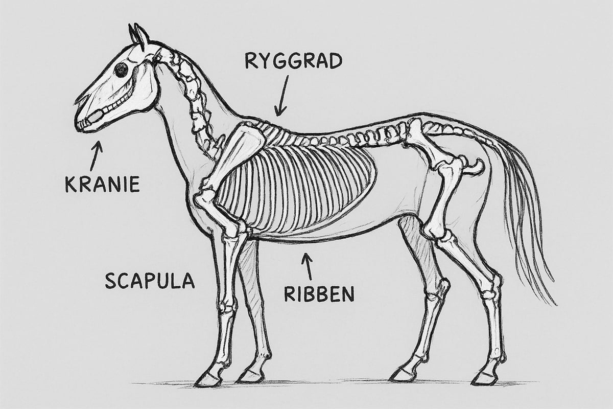
An adult horse skeleton typically consists of about 205 individual bones. Together, these bones form an advanced structure that can be divided into four main parts: the skull, spine, ribs, and fore and hind limbs. The horse skeleton differs from the human skeleton in having a more elongated spine and stronger limbs, which is necessary to support the horse's weight and enable rapid movement.
The main functions of the horse skeleton include:
- Support for the body and muscles
- Protection of internal organs
- Creation of movement via joints and muscles
A special feature of the equine skeleton is the strong and flexible spine, which allows natural movements during riding and work. For example, an average adult horse has a bar measurement of approximately 150 cm, which reflects the proportions and strength of the skeleton. For more in-depth information on structure and function, you can read Anatomy and Physiology of Horses .
The horse skeleton is designed to withstand significant loads, and it is this foundation that makes the horse one of the most versatile mammals.
Structure and Function of the Skull
The skull in the horse skeleton plays a central role in protecting the horse's brain and sensory organs. The robust skull encloses the brain and forms a framework for the eyes, ears and nostrils. The tooth structure is also part of the skull and is crucial for how the horse absorbs and chews its food.
There is great variation in skull shape between breeds, which affects both appearance and function. For example, some breeds may have longer muzzles than others. A notable anatomical feature is the so-called bolt hole, which is used in veterinary practice during euthanasia to ensure correct placement.
The skull in a horse skeleton clearly shows how evolution has adapted the shape and function of the bones to the horse's needs.
Spine and Ribs: Stability and Protection
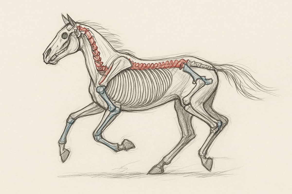
The spine in the equine skeleton consists of several sections: cervical vertebrae, thoracic vertebrae, lumbar vertebrae, sacrum, and coccyx. Together, these vertebrae form a flexible but stable column that must be able to both bend and withstand load during movement. The number of vertebrae may vary slightly between individuals, but the structure is basically the same.
The ribs protect the internal organs such as the heart and lungs. They are connected to the thoracic vertebrae and form a solid but slightly flexible frame. The balance between flexibility and stability in the spine is crucial for the horse to move freely without compromising its health.
The horse's skeleton ensures that vital organs are well protected, while the spine allows the necessary movements for all gaits.
Forelimbs and Hindlimbs: Movement and Load
The forelimbs of the horse skeleton consist of the scapula, humerus, radius, and carpus. These bones work together through joints such as the shoulder, elbow, and knee joints, enabling the horse to absorb shock and move efficiently.
The hind limbs include the pelvis, femur, tibia, and tarsus. The joints in the hind limbs, including the hip and hock joints, are essential for power transmission and propulsion. The structure of the hoof is also unique to the equine skeleton and acts as a natural shock absorber that protects the rest of the leg from injury.
Different types of joints contribute to mobility and strength, enabling the horse to perform both fast and powerful movements. The horse's skeleton is thus optimized to handle both weight and movement in all situations.
Functions and Role of the Horse Skeleton in Movement
The equine skeleton is the foundation for the horse's ability to move efficiently, safely and elegantly. It enables everything from calm strides to explosive leaps, and each bone plays a crucial role in the overall interplay between strength, flexibility and control. To understand the horse's movement potential, one must delve into the different functional aspects of the equine skeleton.
The role of the skeleton in the horse's motor skills
The horse's skeleton forms the foundation for all of the horse's movements. For example, it enables different gaits such as walk, trot and gallop, where bones, joints and muscles work closely together. A well-functioning horse skeleton ensures that the horse can optimally utilize its strength and agility.
When the horse moves, power is transferred from the hind legs through the pelvis and spine to the forelegs. This power transfer depends on all joints and bones working harmoniously. A galloping horse often has a lighter and more flexible horse skeleton than a working horse, which makes it faster and more agile.
Correct structure of the horse's skeleton is crucial for performance and durability. Irregularities or misalignments can lead to injuries and reduced performance. Therefore, understanding the horse's skeleton and its role in motor skills is essential for anyone who trains, rides or breeds horses.
Joints and Mobility
The joints in the horse's skeleton are designed to provide both strength and mobility. The three primary types of joints are ball joints, hinge joints, and gliding joints, each of which allows for different movements. Ball joints allow for rotation, hinge joints provide flexion and extension, and gliding joints allow for small adjustments.
Articular cartilage is an important component as it reduces friction and protects the bones from wear and tear. Wear and tear on the cartilage can lead to pain and limited movement, which is often seen in sports horses. Statistics show that joint injuries are among the most common problems in active horses.
For a more detailed explanation of joint types and their function in the equine skeleton, you can read Skeletal Anatomy: A Thorough Explanation .
Restrictions in mobility are often due to either wear and tear or congenital variations. Regular training and maintenance of the horse's joints is therefore essential to maintaining a healthy horse skeleton.
Support, Protection and Weight Bearing
The horse's skeleton not only enables movement, but also supports and protects the body's vital organs. The ribs form a solid cage around the heart and lungs, while the spine provides both flexibility and stability.
The legs are reinforced with strong bones, tendons and ligaments, so they can withstand the heavy loads of jumping and running. The structure of the hoof plays a special role as shock absorber, protecting the rest of the horse's skeleton from overload.
The table below summarizes important support functions:
| Structure | Function |
|---|---|
| Rib | Protection of organs |
| Backbone | Stability and flexibility |
| Leg | Weight bearing and movement |
| Hoof | Shock absorption |
These adaptations make the horse skeleton uniquely suited for both everyday use and extreme performance.
Development and Adaptation Throughout Life
The horse's skeleton undergoes significant changes from foal to adult. In the first years of life, bones grow rapidly, and the growth plates do not close until late in the young horse's life. This means that young horses are particularly vulnerable to growth-related injuries if they are stressed too early.
As a horse ages, its skeleton adapts to the stress and training it is exposed to. Training programs can strengthen bones and increase their resilience, while inactivity can lead to weakening. Bone strength often peaks in the middle of a horse's life and gradually declines.
It is therefore important to adapt training to the horse's age and stage of development to ensure a healthy horse skeleton throughout its life.
Special Anatomical Adaptations
The horse's skeleton has evolved to support speed and endurance. The elongated spine and strong, slender legs allow the horse to move quickly over long distances.
Unique features, such as the structure of the forelimbs without collarbones and specialized joints, distinguish the horse skeleton from other mammals. These adaptations make the horse one of the most efficient land animals when it comes to running and locomotion.
Common Problems and Diseases of the Horse Skeleton
The equine skeleton is the foundation of a horse's health, but it is also susceptible to various problems and diseases. Understanding the most common challenges is essential to ensuring your horse's well-being, whether you are a rider, owner or professional.
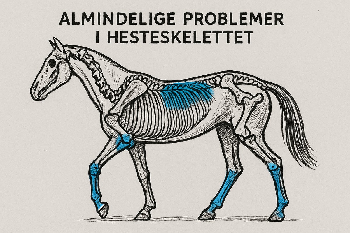
Common Skeletal Injuries and Causes
Injuries to the horse's skeleton often occur as fractures, joint injuries and osteoarthritis. These problems typically occur from overuse, falls or accidents during training and competition. Genetic predispositions may also play a role, especially in breeds with a tendency towards weak joints.
Statistics show that leg fractures are one of the most common serious injuries in riding horses, and the risk increases with the intensity of use. Fractures are particularly common in the forelegs, where weight bearing is greatest.
Typical causes:
- Overexertion during jumping or galloping
- Trauma from a fall or collision
- Hereditary factors
For more information about research on musculoskeletal disorders in animals, read about research on musculoskeletal health .
Diseases and Degenerative Conditions
The horse's skeleton is often affected by diseases such as osteochondrosis, arthritis and bone cysts. Osteochondrosis is caused by disturbances in bone development and is especially seen in young horses. Arthritis, or osteoarthritis, develops gradually and leads to pain and reduced mobility.
Symptoms of equine skeletal disease include lameness, swelling, and altered gait. Diagnosis is often made through clinical examination and imaging such as X-rays or ultrasound.
The importance for the horse's quality of life is great, as even mild degenerative changes can affect performance and well-being. Early detection is therefore crucial to reduce the consequences.
Prevention and Maintenance
Preventing problems with the horse's skeleton requires a holistic approach. Proper exercise, adapted to the horse's age and development, strengthens bones and joints. Feed with the right content of calcium, phosphorus and vitamin D is vital for optimal bone development.
Regular health checks make it possible to detect incipient injuries or diseases of the horse's skeleton early. Training programs should include varied terrain and gradual escalation of load.
To summarize:
- Balanced feeding plan
- Customized training and rest
- Routine health check-up
Treatment and Rehabilitation
Treatment for equine skeletal injuries ranges from conservative approaches to advanced surgery. Minor injuries are often treated with rest, bandaging, and physical therapy, while serious fractures may require surgery.
Medical treatment includes painkillers and anti-inflammatory drugs. Rehabilitation is a gradual process where the horse slowly retrains after the injury. The success rate of treatment for skeletal injuries depends on the type of injury and timely intervention.
Examples of successful rehabilitation include the use of specialized training programs where the horse's skeleton is gradually strengthened and the risk of relapse is minimized.
Modern Teaching and Anatomical Models
Modern teaching of the equine skeleton has undergone significant development in recent years. New teaching methods and advanced models make it easier to understand complex anatomical relationships. This section delves into how physical and digital resources are used in teaching, and how ethics, quality and innovation play a central role.
The Importance of Anatomical Models in Learning
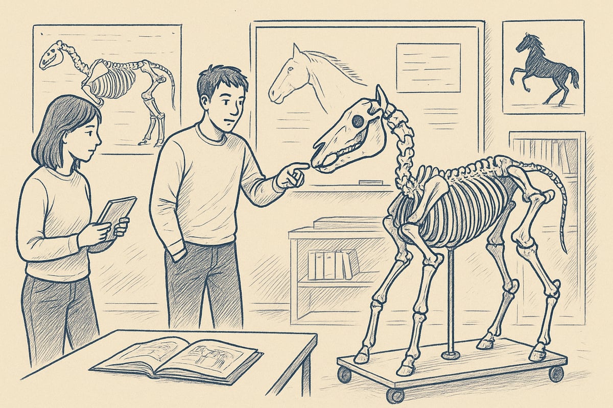
Physical models of the horse skeleton are of great importance for learning. Students and teachers have the opportunity to examine the mutual location and structure of the bones in practice. This creates a concrete understanding that is often difficult to achieve through books alone.
Compared to human skeletons, horse skeleton models are often larger and more complex, especially in the structure of the limbs. Using plastic models provides flexibility and durability, while real skeletons provide the most accurate experience. Regardless of choice, the tactile element enhances the learning process.
Digitization and New Learning Methods
Digital developments have revolutionized the teaching of the equine skeleton. 3D models and virtual platforms make it possible to study the bones from all angles and simulate movements. This increases the understanding of both structure and function.
Virtual resources can be individually adapted, making them ideal for both classroom teaching and self-study. Statistics show a significant increase in the use of digital learning tools among veterinary students since the 2020s. This combines the best of physical and digital teaching methods.
Ethics and Quality in the Production of Skeleton Models
Ethical considerations are crucial when making horse skeleton models. Real bones are responsibly collected, often from naturally deceased animals and in collaboration with donors. Processing can take several months to ensure both hygiene and preservation of detail.
The quality requirements are high, especially for educational use. The models must be anatomically correct and durable, so they can be used again and again. This ensures that students and professionals get the most out of their equine skeleton education.
Practical Examples and Application
Horse skeleton models are widely used in clinical practice, research and teaching. Mobile models on a stand with wheels make it easy to demonstrate the function of the bones in different situations. This provides a flexible and effective learning environment.
Storage and maintenance are important so that the models maintain their quality over time. They can be easily transported between classrooms, making them particularly suitable for workshops and courses on the equine skeleton.
Anatomical Posters and Visual Aids
Visual aids such as anatomical posters provide a quick overview of the horse skeleton and its structure. Popular posters often measure 70x100 cm and are used in both teaching and clinical practice. They clearly illustrate bones, joints and proportions.
The advantage of posters is that they can be hung up and used as a reference in daily practice. Many educators choose to supplement models with posters to enhance visual learning. Read more about the use of anatomy posters in teaching and their role in understanding the equine skeleton.
eAnatomy: Anatomical Models and Posters for Teaching
eAnatomi ApS offers a comprehensive range of models and posters covering the horse skeleton in detail. The collaboration with healthcare specialists ensures that each product is both scientifically accurate and of high quality.
The products are used internationally by students, educators and veterinarians. Special measurements and individual solutions are possible. Both physical and digital resources are available, making it easy to adapt the teaching. Read more about the company's expertise atAbout eAnatomy and our work .
Developments in Equine Anatomical Research and Future Perspectives
Research into the equine skeleton is rapidly evolving and constantly bringing new insights that benefit both the health and performance of horses. Modern technologies and interdisciplinary collaborations have made it possible to understand the equine skeleton at a more detailed level than ever before. This knowledge will have great importance for the future education, breeding and daily handling of horses.
Latest Research and Technological Advances
In recent years, research into the equine skeleton has focused on biomechanics and advanced imaging. Using CT and MRI scans, researchers can now examine the microstructure of bones and detect even small changes. Genetic analyses reveal hereditary predispositions to skeletal diseases, enabling early intervention and targeted breeding.
Research shows that proper horse skeletal structure is crucial for both performance and injury prevention. Comparative studies between breeds and types of use provide a deeper understanding of how the horse skeleton adapts to different loads. Readers can find more in-depth articles and reviews of new models and technologies in We Go Deep with Product Writing and Reviews .
Future Skeleton Models and Teaching
Teaching about the equine skeleton is moving towards more interactive and digital solutions. The development of 3D models, augmented reality (AR) and virtual reality (VR) makes it possible to explore the equine skeleton virtually and from multiple angles. This strengthens the understanding of complex structures and facilitates the acquisition of knowledge for both students and professionals.
The need for flexible, personalized learning resources is growing. Many institutions now use a combination of digital resources and physical models. For more information about resources for teaching and students, visit For Students and Educators .
Practical Application in the Horse World
The application of new knowledge about the equine skeleton is clearly visible in modern breeding, training and healthcare. Research is transferred directly into practice, where elite horses are monitored with advanced imaging diagnostics, and management is adapted to individual needs. A concrete example is the use of CT scanning to assess joints and bones before major competitions.
Collaboration between researchers, veterinarians and trainers ensures that the latest knowledge about the equine skeleton is implemented effectively, enhancing both the horse's welfare and performance in the long term.
Knowledge for Owners, Riders and Professionals
Understanding the equine skeleton is important for anyone who works with horses, whether you are an owner, rider or professional. Knowledge of structure and function helps prevent injuries and improve daily handling.
There are many resources and courses that delve into the subject of the equine skeleton and its significance. Investing in knowledge pays off for both horse and human.
Once you have gained insight into the fascinating skeletal structure of the horse and the many functions that support both movement and health, it can be valuable to delve further. Whether you are a student, teacher, veterinarian or just a curious horse owner, anatomical models and visual aids can make your understanding even stronger. At eAnatomi you will find a wide selection of quality-assured skeletal models and posters, developed in collaboration with experts, so you always have access to the latest knowledge and the best learning tools.
Read more here
0 comments