
Did you know that the skin is the body's largest organ, covering about 2 m² in an adult? Many people overlook how complex the anatomy of the skin actually is. In this guide, you will get an in-depth, updated insight into the anatomy, layers and functions of the skin with a focus on 2026.
Whether you work in the healthcare sector, are interested in beauty, or are studying biology, knowledge of skin anatomy is essential. The structure of the skin has a major impact on everything from health and aging to general well-being.
Read on to become an expert on the skin's layers, functions, cell types, common skin problems, and the latest research in the field.
The Overall Structure and Function of the Skin
The anatomy of the skin is far more complex than most people realize. In an adult, the skin typically covers about 2 square meters and weighs up to 4 kilograms, making it the largest organ in the body. This multi-layered organ does not simply function as an outer covering, but is made up of sophisticated structures, each of which is crucial to our health and well-being.
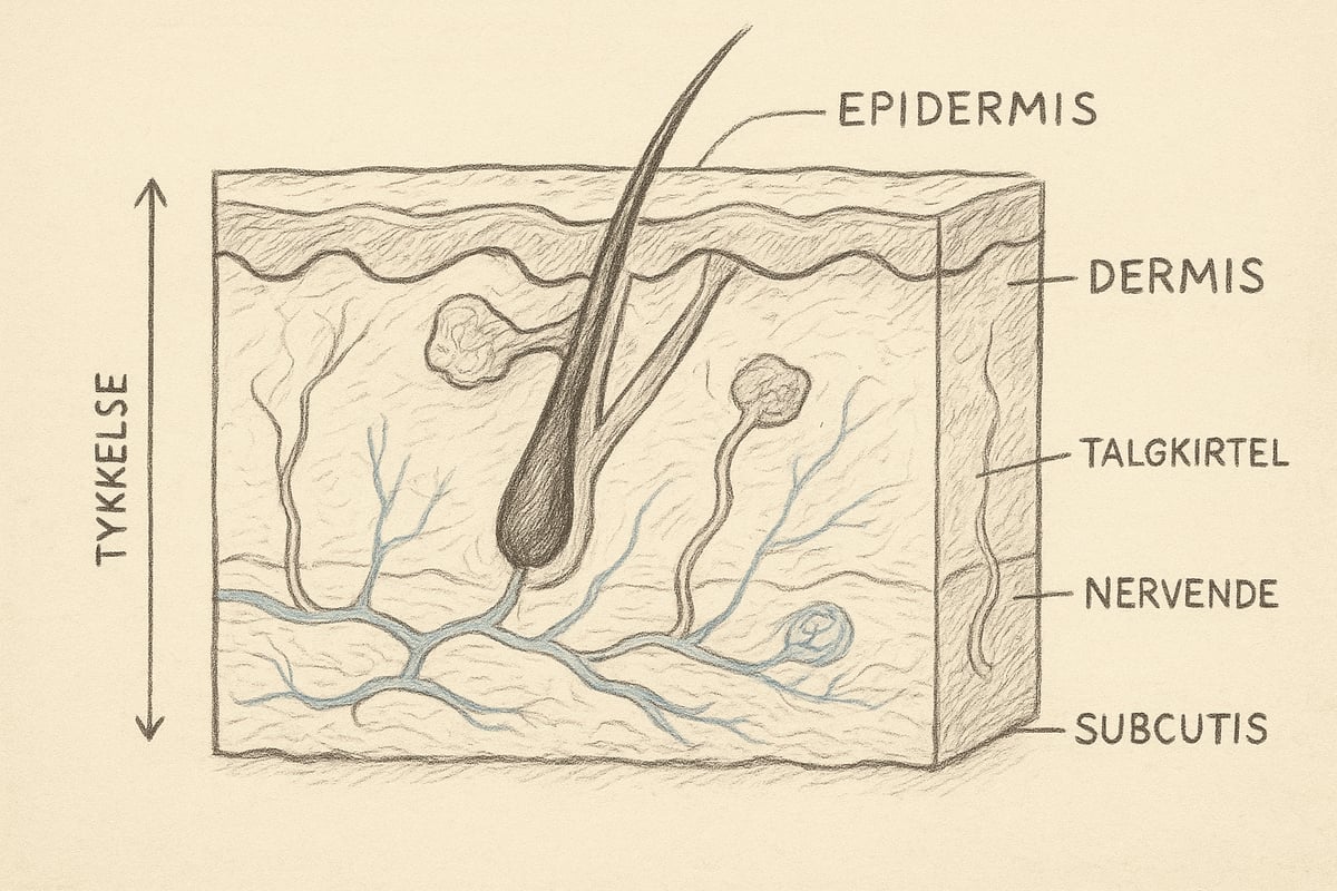
The three main layers of the skin: Epidermis, Dermis and Subcutis
The anatomy of the skin consists of three main layers: the epidermis, the dermis, and the subcutis. Each layer has unique properties and varies in thickness depending on body region and function. For example, the epidermis is only about 0.05 mm thick on the eyelids, but can reach 1.5 mm on the palms of the hands and soles of the feet. The dermis is significantly thicker and contains most of the skin's structures, while the subcutis is primarily composed of adipose tissue and serves as insulation and energy storage.
Transition zones between the layers ensure that nutrients, signaling molecules and cells can move appropriately. The total surface area of the skin provides space for millions of sensory cells and glands. The weight of the skin typically constitutes 6-16% of the body weight, which emphasizes its physiological importance.
| Skin layer | Thickness (mm) | Function |
|---|---|---|
| Epidermis | 0.05 - 1.5 | Barrier, pigment |
| Dermis | 0.3 - 4.0 | Support, senses |
| Subcutaneous | 1.0 - several cm | Insulation, energy |
This complexity in the skin's anatomy is the basis for its many functions and ability to protect the body.
Functions of the skin
The anatomy of the skin enables a number of vital functions that we often take for granted. First and foremost, the skin acts as an effective barrier against microorganisms, chemicals and UV radiation. This protects the body from infections and harmful external influences.
The skin also regulates body temperature through sweat production and adjustment of blood flow in the superficial blood vessels. When the body needs to cool down, sweat production increases, while the blood vessels dilate to release heat.
As a sensory organ, the skin is equipped with a dense network of nerves that detect touch, pressure, pain, heat and cold. This allows us to respond quickly to environmental influences.
Another important function is the synthesis of vitamin D, which is activated when the skin is exposed to sunlight. This vitamin is important for bone health and the immune system.
In addition, the skin helps maintain the body's water balance and protects against fluid loss. Sweat glands help excrete waste products, emphasizing the skin's role in the body's cleansing system.
In severe injuries such as burns or certain diseases, several of these functions can be weakened. Loss of barrier function can lead to infections and fluid loss, while reduced ability to regulate temperature can threaten vital processes.
For an even deeper understanding of how the anatomy and functions of the skin are related, you can read more at Skin Anatomy and Functions .
Epidermis: Structure, Cell Types and Regeneration
The epidermis is the outermost layer of the skin's anatomy and plays a central role in protection, color, and renewal. This layer is fascinating because it constantly renews itself and contains several cell types, each of which has a crucial function for skin health. Understanding the structure and processes of the epidermis is essential for anyone who wants deep insight into the anatomy of the skin.
Structure and layering of the epidermis
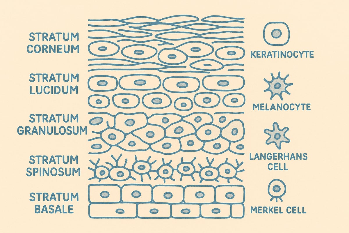
The epidermis consists of five layers, which together form the outermost protective layer of the skin's anatomy. The five layers are:
- Stratum corneum (corneal layer): Consists of dead, keratinized cells that form a strong barrier.
- Stratum lucidum: Found only in thick skin, such as on the palms of the hands and soles of the feet.
- Stratum granulosum: Contains cells that begin to die and fill with keratin.
- Stratum spinosum: Consists of several layers of keratinocytes with strong connections.
- Stratum basale: The deepest layer where cell division takes place.
The thickness of the epidermis varies considerably depending on its location on the body. On the eyelids it is only about 0.05 mm, while on the palms of the hands and soles of the feet it can be up to 1.5 mm. The transition zones between the layers ensure that the skin is both flexible and strong.
The main cell types in the epidermis are:
| Cell type | Function |
|---|---|
| Keratinocytes | Forms keratin, protects against the environment |
| Melanocytes | Produces melanin, controls skin color |
| Langerhans cells | Immune defense against foreign substances |
| Merkel cells | Sensory function, detects touch |
If you want to delve further into the layers, you can read more at Skin Anatomy and Layers , which reviews the structure in detail.
Melanin and skin color
Melanocytes in the epidermis produce the pigment melanin, which is essential for skin color and protection. Melanin comes in two main types: eumelanin (dark brown/black) and pheomelanin (red/yellow). The distribution of these pigments determines individual skin color.
Skin anatomy varies globally, and the amount and type of melanin have a major impact on UV protection. People with high eumelanin production have better natural protection against the sun's harmful rays, while people with more pheomelanin are more vulnerable to UV damage.
Melanocytes are scattered throughout the stratum basale and transfer melanin to surrounding keratinocytes. This process is essential for protecting the deeper layers of the skin from DNA damage caused by ultraviolet light.
Variations in skin color are a result of both genetic factors and adaptation to sunlight through evolution. As a result, there are large differences in skin anatomy and pigmentation across the world.
Epidermis regeneration and wound healing
One of the most remarkable properties of the epidermis is its ability to regenerate. Stem cells in the stratum basale ensure the constant formation of new keratinocytes, which slowly move upwards through the layers. This entire process, called keratinization, takes approximately 28 days.
The epidermis is renewed approximately every four weeks in adults. This rapid cell turnover is essential for maintaining the skin's anatomy and protecting it from external influences. Damage to the epidermis, for example from wounds, activates a complex healing process in which stem cells migrate to the wound area and form new tissue.
Disruptions in the regeneration process can lead to skin conditions such as psoriasis or eczema, where cell production is either too fast or too slow. This affects both the skin's structure and its ability to heal effectively.
The rate of wound healing depends on several factors, including age, nutrition, and the presence of diseases, emphasizing the importance of a healthy epidermis for the entire skin anatomy.
The skin's immune defense
The epidermis contains specialized cells, particularly Langerhans cells, which play a key role in the skin's immune defense. These cells act as "sentinels" that capture foreign substances and present them to the immune system.
When the skin is exposed to allergens or infectious microorganisms, Langerhans cells are activated and initiate an immune response. This is central to protecting the body against bacteria, fungi and viruses that could otherwise penetrate the skin barrier.
The skin's anatomy makes the epidermis the first line of defense against external threats. In diseases such as atopic eczema or allergic reactions, an overreaction of the immune system is often seen, which can lead to redness, itching and inflammation.
A well-functioning immune defense in the epidermis is therefore crucial for skin health and the body's overall protection. Understanding these mechanisms provides insight into why the anatomy of the skin is so vital to our well-being.
Dermis: Support Structure, Collagen and Skin Elasticity
The dermis is the middle layer of the skin's anatomy and is the foundation for the skin's strength, elasticity and sensitivity. This layer is much thicker than the epidermis and forms the framework for many of the skin's most vital functions. To understand how the skin protects, senses and remains supple, it is essential to know the structure and role of the dermis.
Dermis layers and composition
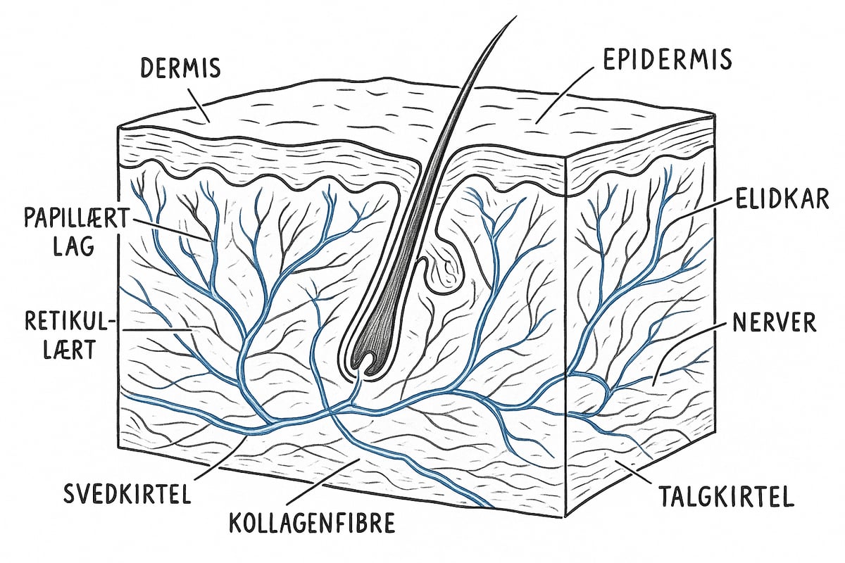
The dermis consists of two main layers: the papillary layer, which is the topmost and rich in thin collagen fibers, and the reticular layer, which is thicker and contains strong, tightly woven collagen bundles. These layers give the skin its strength and flexibility.
Connective tissue dominates the dermis and consists of collagen and elastin fibers, which ensure that the skin can both resist tension and return to its original shape. This layer also contains blood vessels, lymphatic vessels and a network of nerves, all of which play a central role in the skin's anatomy.
The thickness of the dermis varies from 0.3 mm in the eyelids to 3 mm on the back. For a visual overview , Anatomical posters about skin and hair can provide a detailed picture of the layers and their relative positions.
The skin's sensory organs in the dermis
The dermis contains several types of sensory organs, making the skin the body's largest sensory organ. Meissner corpuscles detect light touch, while Pacinian corpuscles respond to vibration and pressure. Pain receptors are also found in the dermis and ensure that the skin can respond quickly to damage.
Sensitivity varies on different parts of the body. For example, fingertips have many Meissner corpuscles, making them very sensitive, while the back has fewer and therefore lower sensitivity. The anatomy of the skin and the location of the sensory organs explain why we perceive stimuli so differently.
These sensory organs work together with the rest of the nervous system to provide detailed information about the environment and ensure rapid responses to heat, cold and pain.
Blood vessels and temperature regulation
The blood vessels in the dermis play a crucial role in the body's temperature regulation. When the body needs to cool down, the blood vessels dilate (vasodilation), which increases blood flow and makes it easier to release heat to the environment. Conversely, the blood vessels contract (vasoconstriction) when cold to conserve heat.
This process is clearly seen when the skin turns red in heat or physical activity, or when we turn pale in cold or shock. The anatomy of the skin allows the body to adapt quickly to changing temperatures and protect the internal organs from heat loss.
The dense network of blood vessels in the dermis is therefore a prerequisite for the body to maintain a stable temperature and respond effectively to external influences.
Skin aging and collagen loss
Aging significantly affects the anatomy of the skin, especially in the dermis. Collagen production decreases by approximately 1 percent per year from the age of 25, gradually making the skin thinner and less elastic. This is seen as wrinkles, sagging skin and loss of firmness.
Elastin fibers also become fewer and more disorganized with age, further reducing the skin's ability to return to its original shape after pressure or stretching. External factors such as sunlight, pollution, and smoking can accelerate this process.
Loss of collagen and elastin are the main causes of visible signs of aging, and understanding these processes is central to both skincare and medical treatments of the skin.
Sweat and sebaceous glands
The sweat glands in the dermis are of two main types: eccrine and apocrine. Eccrine glands are distributed throughout the body and regulate temperature by secreting sweat, while apocrine glands are found mainly in the armpits and groin and are activated by emotional stimuli.
Sebaceous glands produce sebum, which keeps the skin supple and protects it from drying out. The balance between sweat and sebum production is essential for the skin's anatomy, as it ensures optimal moisture and protection.
Disturbances in the functioning of the glands can lead to problems such as dryness, acne or excessive sweating. The dermis' role in housing these glands highlights how complex and adaptable the skin is.
Subcutis: Adipose Tissue, Insulation and Energy Storage
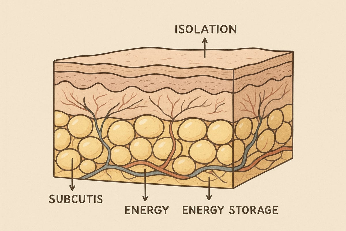
The subcutis is the deepest layer of the skin's anatomy and acts as the body's natural cushioning. This layer consists primarily of fat cells, called adipocytes, which form a soft but robust network beneath the dermis. The thickness of the subcutis varies significantly from person to person and depends on both genetics, gender and body region. For example, the subcutis can be a few millimeters thick on the eyelids, while around the abdomen or thighs it can measure several centimeters. This variation is central to the anatomy of the skin, as it affects both the appearance and function of the body.
The subcutis plays a number of important roles in the body's daily functioning. First and foremost, the layer provides insulation, allowing the body to retain heat, even in cold conditions. In addition, the subcutis acts as a shock-absorbing layer, protecting muscles and organs from impact and pressure. The adipose tissue also acts as an energy store that can be mobilized during periods of increased need. An overview of the functions of the subcutis can be summarized in the table below:
| Function | Description |
|---|---|
| Insulation | Protects against heat loss |
| Shock absorption | Absorbs impact and pressure |
| Energy storage | Stores energy in the form of fat |
| Design | Affects body contours |
Together with the other layers of the skin's anatomy, the subcutis forms an indispensable part of the body's protection and energy management.
When we look more closely at the role of the subcutis in the body's metabolic health, its importance becomes even clearer. The fat cells in the subcutis produce, among other things, the hormone leptin, which regulates appetite and energy expenditure. The distribution of adipose tissue in the subcutis varies between individuals, and this distribution has a major impact on both BMI and the risk of diseases such as type 2 diabetes and cardiovascular disease. An uneven or excessive fat distribution can increase health risks, while a balanced subcutaneous fat layer supports the body's normal functions.
The anatomy of the skin therefore includes not only the visible layers, but also the deeper structures that influence the well-being of the entire body. Understanding the functions of the subcutis is essential for assessing the health of the body, both in clinical practice and in everyday life. Precisely because the subcutis constitutes a large part of the skin anatomy, it is important to be aware of how lifestyle and genetics can affect this layer.
A particularly important function of the subcutis in the anatomy of the skin is its role in wound healing. The subcutis contributes blood supply and delivers nutrients to the overlying layers, which promotes the healing process after injuries or surgeries. However, with low subcutaneous fat, healing can be slower because there are fewer resources available for tissue repair. This is often seen in people with low body fat percentage or certain medical conditions.
If you want to delve deeper into how the subcutis and the other layers work together during wound healing, you can read more at The Role of the Skin in Wound Healing , where you will find a detailed review of the layer's importance in the healing process. The subcutis is therefore not only a depot for energy, but also an active participant in the body's defense and repair.
Skin Types, Skin Problems and Modern Skin Research
Different skin types and their characteristics
The anatomy of the skin varies from person to person, and so do our skin types. Knowing your skin type is essential for proper skin care and understanding how your skin reacts to environmental influences. The most common skin types fall into five categories: normal, dry, oily, combination, and sensitive skin.
| Skin type | Characteristics |
|---|---|
| Normal | Balanced, rarely impurities |
| Dry | Tight, scaly, easily irritated |
| Greasy | Shiny, acne prone |
| Combined | Oily T-zone, dry cheeks |
| Sensitive | Mild redness, itching, reactions to products |
According to recent research, skin types are distributed differently in the population, but sensitive skin is experienced by up to 50%. The anatomy of the skin determines how these types arise, as sebum production, moisture balance and barrier function are controlled by the skin's layers and cells.
Common skin diseases and conditions
Skin anatomy plays a central role in the development of skin diseases. Acne, for example, affects around 80% of adolescents and is caused by overproduction of sebum and inflammation in the skin's layers. Psoriasis, rosacea and atopic eczema are other common conditions that affect both appearance and quality of life.
Symptoms vary: Acne appears as pimples and blackheads, while psoriasis causes scaly, red patches. Rosacea causes redness and visible blood vessels, and atopic eczema manifests as itchy, dry skin.
For a visual overview of typical skin disorders and the structure of the skin, you may benefit from viewing this Skin Anatomy and Disorders poster , which illustrates the connection between structure and disease.
The skin's reaction to environment and lifestyle
The anatomy of the skin makes it extremely sensitive to external influences. UV radiation, pollution, diet and stress all have a direct impact on the appearance and health of the skin. Too much sun can lead to premature aging, pigment changes and increase the risk of skin cancer.
Pollution contributes to inflammation and can worsen existing skin problems. Unhealthy diets and stress can weaken the skin barrier and lead to acne or eczema outbreaks. The skin's anatomy determines how resistant the skin is to these influences, as thickness, pigmentation and cell turnover vary.
Modern skin research and technological advances
The field of skin anatomy has seen significant advances in recent years. New treatments such as laser, microneedling, and biotechnological solutions improve both aesthetics and function. Stem cell research and regenerative medicine offer hope for patients with serious skin diseases or injuries.
The market for skincare and dermatological treatments is expected to grow by 5% annually until 2026. This is due to increased focus on scientifically documented methods and tailored solutions based on the skin's anatomy and individual needs.
Skin anatomy in teaching and practice
A thorough understanding of skin anatomy is essential for medical students, cosmetologists, and healthcare professionals alike. Teaching about the skin's layers, cell types, and functions is often done with anatomical models and posters that visualize complex relationships.
In practice, this knowledge is used to diagnose skin diseases, guide skin care and develop new treatments. If you want to delve deeper into learning material about the anatomy of the skin, you will find a wide selection of teaching material about anatomy , which can strengthen both theoretical and practical understanding.
As we delve into the fascinating world of the skin, we discover how complex and important this organ is to our health, well-being and daily life. Whether you work in healthcare, study anatomy or are simply curious about how your skin works, this guide will give you a solid foundation for understanding the layers and functions of the skin. If you want to explore even further, see anatomical models or find visual posters that can strengthen your knowledge or teaching, you can get inspiration and find the right tools here:
Read more here
0 comments