

Understanding the structure and function of the body becomes much more tangible when we use anatomical models in teaching and healthcare contexts. These models are indispensable for students, educators, and professionals alike, as they create visual clarity and practical insight.
This article will surely guide you through everything you need to know about anatomy models in 2026. You will gain knowledge about selection, use, the latest trends and a detailed buying guide, so you are well equipped to make the best choice.
We cover the different types of models, their applications, materials, selection criteria, upcoming trends, maintenance and where to find the best ones on the market. The goal is to give you a complete overview, whatever your needs.
Visual learning, technological advances, and the increased use of realistic models have made anatomy models central to modern education and healthcare. Read on to discover how you can get the most out of them in your work or study.
What are anatomy models?
Anatomy models play a central role in understanding the structure and function of the body. They enable us to experience anatomy in a concrete way, which enhances both learning and communication. But what does the term actually cover, and why are these models so important in education, healthcare and research?
Definition and Purpose
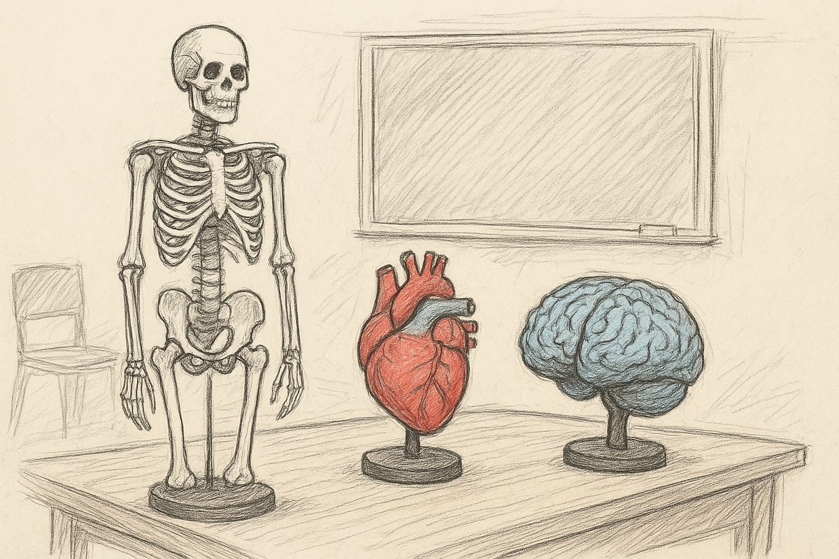
Anatomy models are physical or digital representations of human or animal anatomy. They are widely used for teaching, demonstration, research, and patient communication. Examples of anatomy models include skeletal models, organ models, and tissue models. The purpose is to make complex structures tangible and easy to understand.
In Denmark, 85% of health education programs use anatomy models in teaching according to Scandidact/Proterapi. Compared to two-dimensional illustrations, these models provide a much deeper understanding and a greater learning outcome. For a comprehensive overview of the many types, see Anatomical models overview .
Historical Development
The development of anatomical models has been significant over time. In the 19th century, wax models were widespread, while today's models range from classic plastic versions to advanced 3D printing and AR solutions. Manufacturers such as 3B Scientific and Somso have set the standard for quality and innovation.
Materials have become more realistic, and digital solutions have opened up simulation training and increased accessibility. Technological advances have made anatomy models more detailed and user-friendly, so that more people can benefit from them in both teaching and practice.
Types of Anatomy Models
There are many types of anatomy models, covering everything from skeletons and organs to joints, tissues and microscopic structures. Examples include the 3B Classic Skeleton, models of brain ventricles and functional joint models.
The types can be categorized by use:
- Education: Basic models for schools and universities
- Clinical Training: Functional Joints and Organs
- Patient information: Clear and illustrative models
- Veterinary use: Animal models for teaching and practice
The benefits of each type depend on the target audience, while limitations often relate to level of detail and price.
Benefits of Using Anatomy Models
The use of anatomy models significantly improves visual and tactile learning. It becomes easier for healthcare professionals to communicate with patients because the models make it possible to clearly explain diagnoses and treatment options.
Repetitive training can be carried out without risk to the patient, which is essential in both education and clinical practice. Studies from Scandidact show that students achieve better exam results when using anatomy models. For example, physiotherapists use joint models to illustrate injuries to patients, which increases understanding and collaboration.
Areas of application for Anatomy Models
Anatomy models play a central role in many different contexts. They are widely used in education, healthcare, research, patient communication and veterinary medicine. By making complex anatomical structures understandable, anatomy models support learning, training and education across disciplines.
Educational institutions
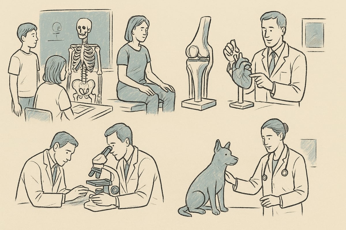
In the world of education, anatomy models are indispensable for creating an understanding of the structure of the body. Elementary schools, high schools and universities use anatomy models to illustrate everything from skeletal structures to internal organs. Especially in medical school and nursing programs, the models provide an opportunity for practical and visual learning.
Over 90% of teachers consider anatomy models to be essential tools in subjects such as biology and anatomy. When complex topics such as the nervous system need to be taught, models make it easier to translate theory into practice. Anatomy models can be adapted to the level of teaching, so that both children and adults get optimal benefit. This versatility makes them a permanent element in modern pedagogy.
Healthcare sector
In the healthcare sector, anatomy models are used daily for both training and patient guidance. Hospitals, clinics, physiotherapists and dentists use anatomy models to improve communication with patients and colleagues. For example, physiotherapists use knee joint models to illustrate rehabilitation plans and injury progression.
Anatomy models help healthcare professionals explain complex procedures in an easy-to-understand manner. This increases patient understanding and compliance, which often leads to better treatment outcomes. The models are also used in preparation for surgeries, where accurate visualization is essential for success.
Research and Development
In research and development, anatomical models are an important tool for testing hypotheses and developing new medical technologies. Scientists use anatomical models to investigate biomechanics, develop prosthetics, and simulate surgical procedures without risk to patients.
By using anatomical models, researchers can visualize and test solutions in a controlled setting. This helps to promote innovation, as errors and improvements can be identified early in the process. In addition, anatomical models play a central role in testing medical devices before clinical use.
Patient Communication and Information
Effective patient communication often requires more than just words. Anatomy models allow doctors and healthcare professionals to show the patient what is happening in the body during a disease or treatment. For example, a heart model can illustrate how a blood clot affects the circulation, making it easier for the patient to understand their diagnosis.
This visual communication strengthens the patient's security and engagement. Anatomy models also facilitate informed consent, as the patient gets a more realistic picture of what an operation or procedure entails. This makes the patient an active participant in his or her own treatment process.
Veterinary Medicine and Specialized Fields
In veterinary medicine, anatomical models are indispensable for both teaching and practice. Animal models are used in veterinary education to train diagnostic and surgical skills. Practicing veterinarians use anatomical models to explain diseases and treatments of pets and farm animals to owners.
Examples range from cattle and horse models to detailed dog anatomy models. To see a wide selection of these models, visit Veterinary Anatomy Models , where various solutions for both teaching and clinical use are presented. The use of anatomy models in the veterinary sector ensures that both professionals and pet owners gain the best possible understanding.
Materials and Technology Behind Modern Anatomy Models
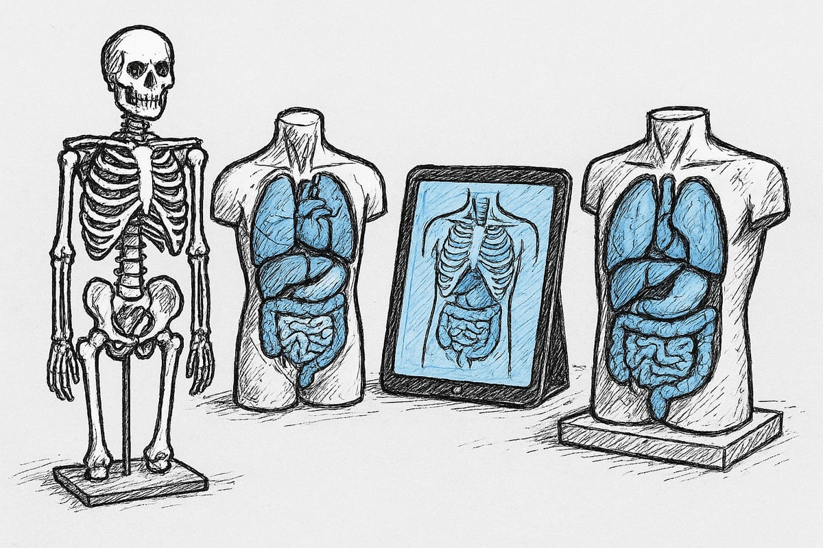
Developments in materials and technology have transformed anatomy models into advanced learning tools. Today's models range from classic skeletons to interactive, digital solutions. The choice of materials and technology has a great impact on both function, durability and learning outcomes.
Classic Materials
Classic anatomy models are often made of plastic, plaster, wax or wood. These materials have been the basis for teaching and demonstration models for many years. Plastic is popular because it is robust and easy to clean. Plaster provides a high level of detail, but is heavy and fragile. Wax was previously used for very detailed models, especially in the 19th century, but requires careful storage.
One of the most widely used anatomy models is the hard plastic skeleton model, which makes it possible to illustrate movements and joints. Wooden models are best suited for rough overviews, but often lack detail.
| Material | Benefits | Disadvantages |
|---|---|---|
| Plastic | Lightweight, durable | May lose color |
| Plaster | Very detailed | Fragile |
| Wax | Realistic | Sensitive to heat |
| Wood | Naturally | Get details |
Classic anatomy models still provide great value in teaching and ensure tactile understanding of the structure of the body.
Advanced Materials and 3D Printing
Today we see a trend towards more advanced materials in anatomy models. Silicone and flexible plastics provide a realistic feel and allow for tissue elasticity to be simulated. Biocompatible materials open up possibilities for use in medical training and surgical simulations.
3D printing has revolutionized the production of anatomical models. 3D printing allows the development of customized models that reflect the anatomy of individual patients. This technology enables rapid production and accurate reproduction of even complex structures.
Advanced materials ensure that anatomy models not only look realistic, but also feel and behave like real tissue.
Digital Anatomy Models and Augmented Reality
Digital solutions have gained traction in education and training. Anatomy models are now available as apps, AR glasses, and virtual labs. These technologies allow for interactive exploration of body structures without the need for physical storage.
For example, with VOKA Anatomy Pro 3D anatomy models, you can explore the human body in detailed, realistic 3D models. This increases understanding and makes learning more engaging.
Digital anatomy models provide flexibility, access to simulation, and the ability to share content across platforms.
Functional and Interactive Models
Functional anatomy models have moving parts, removable organs or can simulate disease states. Examples are 12-part knee joints or torsos where organs can be removed and examined in more detail.
These interactive anatomy models engage both students and professionals because they provide the opportunity to practice practical skills over and over again. This can be crucial for understanding complex processes and enhances learning through hands-on experience.
Functional models make it easier to translate theory into practice in both teaching and clinical training.
Quality Standards and Certifications
When choosing anatomy models, it is important to ensure that they meet relevant quality standards. CE marking and ISO certification are examples of requirements that document safety and accuracy.
Pedagogical approval ensures that the models are suitable for educational use. Manufacturers such as 3B Scientific and Somso are known for high quality and precision.
By choosing certified anatomy models, you get both security and optimal learning value, regardless of whether it is for education, the healthcare sector or research.
How to Choose the Right Anatomy Model – Step-by-Step Guide
Choosing the right anatomy models requires insight and systematicity. With this step-by-step guide, you will get a clear overview so you can safely navigate through the options and make the best choice for your specific needs.
1. Define Your Purpose
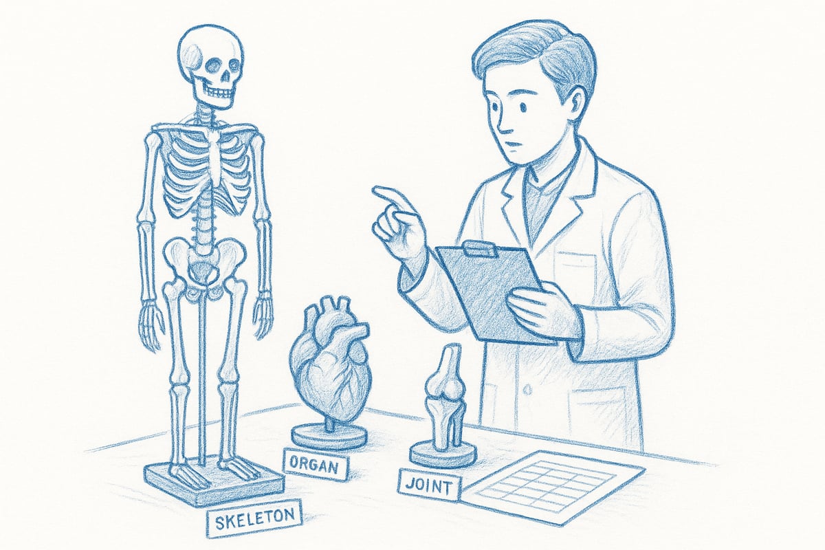
Before choosing from the many anatomy models, you need to clearly define the purpose. Will the model be used for teaching, clinical demonstration, research or patient information? The purpose determines the level of detail and features required.
For example, elementary school education often requires more robust and simple models, while university level or clinical use requires a higher level of detail. A model to be used for patient communication should be intuitive and easy to understand.
By starting from the purpose of your anatomy models, you ensure that the investment provides optimal learning or communication. This makes it easier to identify relevant models and exclude those that do not match the need.
2. Choose the Correct Type and Size
The next step is to choose the right type and size of anatomy models. Consider whether you need a skeleton, organ, joint, tissue or perhaps a microscopic model. Sizes range from small tabletop models to lifelike 1:1 reproductions.
For biology teaching, a classic skeleton model may be an obvious choice, while torso models with removable organs are ideal for medical training. If you want inspiration, you can see a wide selection ofskeleton models for teaching .
The size depends on the space and user group. Smaller models are suitable for classrooms, while larger models are often used in lecture halls or clinical environments. Remember to choose the type of anatomy model that best supports your teaching or training situation.
3. Consider Material and Durability
The choice of material has a great impact on both the usability and longevity of anatomy models. Plastic is lightweight, durable and easy to clean, while silicone provides a more realistic feel, especially when simulating tissues and organs.
Consider how often and how intensively the model will be used. For daily use in teaching or clinical settings, you should choose robust anatomy models that can withstand frequent handling and cleaning. For specialized training, flexible materials can be an advantage.
Investing in quality materials can pay off in the long run, as durable anatomy models require replacement less often. Also consider cleaning requirements and any allergens in the materials to ensure safe use.
4. Examine Functionality and Interactivity
Functionality is a key factor when choosing anatomy models. Consider whether the model should have moving parts, removable organs, or the ability to simulate disease states. Interactive models promote engagement and hands-on understanding.
For example, functional joint models allow for the demonstration of movements, while torsos with removable organs make it easier to explain complex relationships. Simulation trainers can be integrated with sensors to provide feedback during training.
By choosing anatomy models with the right functionality, you can adapt your teaching or training to make it more vivid and effective. This increases both motivation and learning outcomes for users.
5. Compare Manufacturers and Prices
The anatomy model market ranges from budget-friendly solutions to high-end brands with high precision. Well-known manufacturers include 3B Scientific, Somso and Erler Zimmer. It is important to compare both quality and price to find the best match.
Below is an overview of manufacturers and price levels:
| Producer | Price range (DKK) | Characteristics |
|---|---|---|
| 3B Scientific | 1,000 – 8,000 | High accuracy |
| Somso | 2,000 – 10,000 | Very detailed |
| Erler Room | 800 – 6,000 | Good selection |
According to Proterapi/Scandidact, most anatomy models range between DKK 800 and DKK 8,000. Compare features, warranties, and reviews to ensure you get the most for your money.
6. Buy Only from Authorized Dealers
The final step is to purchase anatomy models from authorized dealers. This ensures that you get warranty, support and proper documentation. Unauthorized suppliers cannot offer the same assurance of quality or service.
Authorized dealers often offer same-day delivery, advice and access to spare parts. Should problems arise, it is easier to get help or exchange the model. Always choose anatomy models from suppliers who can document the origin and quality of the product.
By following this guide, you minimize risk and ensure that your anatomy models meet both professional and practical requirements.
Trends and Developments in Anatomy Models 2026
Developments in anatomy models are accelerating rapidly in recent years. New technologies and materials are creating opportunities that seemed unthinkable just a few years ago. Here you will get an overview of the most important trends that will shape the anatomy model market towards 2026.
3D Printing and Customization
3D printing has revolutionized the production of anatomical models. Today, models can be quickly produced that are tailored to specific patients or educational needs. This means that healthcare professionals can plan and practice surgical procedures on models that accurately match the patient's anatomy.
The advantage of this development is both rapid production and high precision. 3D printed anatomical models make it possible to visualize rare or complex conditions that would otherwise be difficult to demonstrate. Several manufacturers now offer customization, allowing institutions and clinics to order models with special features or sizes.
This trend makes anatomy models more relevant in both research and practice.
Digitization and Virtual Models
Digitalization is playing an increasingly important role in the development of anatomy models. AR and VR have made it possible to create virtual laboratories where students can explore the body interactively without physical limitations.
Virtual models can simulate movement, disease states, and surgical procedures. According to a study, 60% of healthcare education institutions expect to increase the use of digital anatomy models by 2026. With AR/VR & 3D printing in anatomy education, educators have access to innovative solutions that combine the best of both worlds.
This development elevates both the learning experience and the availability of advanced knowledge.
Sustainability and Eco-Friendly Materials
Sustainability has become a central theme for anatomy model manufacturers. Several companies have switched to bioplastics and recycled materials to reduce their environmental footprint. This development is happening both for the sake of the climate and to meet the requirements of institutions for green procurement.
The choice of environmentally friendly materials has a major impact on the future procurement of anatomy models. For users, it means that sustainability can be prioritized without compromising on quality or functionality.
Environmentally friendly solutions are expected to become an important selection criterion in the coming years.
Integration with Simulation and Skill Trainers
A notable trend is the integration of anatomy models with simulation and skill trainers. Modern models are often equipped with sensors that can provide feedback on practical exercises. This allows students and professionals to practice procedures realistically and receive objective evaluation of their skills.
The combination of classic anatomy models and advanced technology provides more effective and engaging learning. For example, torsos with built-in sensors are used in CPR training or surgical procedures.
This development significantly strengthens the practical application of anatomy models.
Maintenance, Storage and Durability
Proper maintenance of anatomy models is essential to ensure both longevity and optimal function. Whether you work in education, healthcare or research, it is important to handle the models correctly throughout their entire life cycle.
Cleaning and Hygiene
Regular cleaning is essential to keep anatomy models in good condition. Always use cleaning agents that are suitable for the model material. Plastic models can often withstand alcohol-based cleaning, which effectively removes bacteria and dirt. Silicone models, on the other hand, require milder, pH-neutral products to avoid damage.
To maintain hygiene, wipe down models after each use, especially if used for patient communication or multi-user training. Frequently used anatomy models should be inspected regularly for damage or discoloration. Be sure to follow the manufacturer's cleaning instructions to avoid unnecessary wear.
Storage and Handling
Proper storage significantly extends the life of anatomy models. Place the models in a location out of direct sunlight, as UV rays can degrade the materials over time. Avoid damp environments, as this can lead to mold or deformation, especially with natural materials.
Use padded stands, boxes or cabinets to protect the models from bumps and scratches. If anatomy models are moved frequently, they should be transported in suitable boxes or bags. Always handle the models with clean hands to minimize grease and dirt on the surfaces. These precautions ensure that the models retain both function and aesthetics.
Repair and Spare Parts
Quality anatomy models can often be repaired, making them a long-term investment. Many manufacturers offer replacement parts such as replaceable joints, organs, or small components. If a model loses a part, you can often order a new one rather than replacing the entire model.
It is an advantage to choose models from reputable suppliers where you can access spare parts and support. For example, torso models and organ manikins often offer the option to purchase additional organs or replaceable parts, which makes maintenance easier. Repairing rather than replacing saves both money and resources, and ensures that your anatomy models are always ready for use.
Where Can You Buy Anatomy Models?
Finding the right place to buy anatomy models is crucial for quality, service and warranty. The selection, prices and shopping experience vary significantly depending on whether you shop online or in a physical store. Here is an overview to help you choose the best solution for your needs.
Overview of Authorized Dealers
When investing in anatomy models, it is recommended to choose authorized dealers. In Denmark, Scandidact and Proterapi are among the best-known suppliers. International webshops also offer a wide range, but it is important to ensure that they are certified.
The advantages of authorized dealers are many. You get a warranty, access to spare parts and professional customer service. Many offer same-day delivery, which is an advantage for institutions and clinics with urgent needs for anatomy models.
For example, Scandidact provides advice on choosing a model, so you can ensure that anatomy models suit your professional level and purpose.
Buying Guide: Online vs. Physical Store
The choice between online shopping and physical stores depends on your preferences and requirements. Online stores often have a larger selection of anatomy models and make it easy to compare prices and specifications. You can order 24/7 and have them delivered straight to your door.
Physical stores, on the other hand, offer the opportunity to see and feel the models, which can be crucial for educators and professionals with specific needs. Personal advice and the opportunity to ask questions is a big plus, especially when choosing advanced anatomy models.
In short, online shopping provides flexibility and a wide range, while the physical store offers security and hands-on experience.
Price levels and Financing
The price range for anatomy models ranges from approximately DKK 800 for simple models to over DKK 8,000 for advanced or specialized variants. The price depends on the type, size and manufacturer. Several retailers offer institutional discounts, leasing agreements or financing options, which can ease the budget for schools and clinics.
Below you will find a table summarizing typical price levels:
| Type | Price range |
|---|---|
| Skeleton model | 1,200 – 6,500 kr. |
| Organ model | 800 – 3,500 kr. |
| Functional joint model | 2,000 – 8,000 kr. |
| Animal model | 1,000 – 5,000 DKK. |
To find the best deal on anatomy models, it is recommended to keep an eye on promotions and contact retailers for any discounts on larger orders.
Once you have gained insight into how anatomy models can enhance teaching, research and patient communication, you will probably understand how important it is to choose the right one. Whether you work in healthcare education, run a clinic or just want to strengthen your anatomical knowledge, it is crucial to find models that match your needs and follow the latest trends. At eAnatomi ApS you get access to quality, innovation and a wide selection, developed together with specialists and adapted to both private and professional use.
Want to dive even deeper and see which models are best for you? Read more here
0 comments