Skip to product information
Bækken m. kønsorganer, kvinde - eAnatomi
1/11

Pelvic model with pelvic floor muscles and genitals, female

SKU: A589IIIM
€163,95
Taxes included. Shipping calculated at checkout.
In stock

Pickup available at Frederiksberg

Usually ready in 4 hours

Pelvic model with pelvic floor muscles and genitals, female

Default Title

Frederiksberg

Pickup available, usually ready in 4 hours

Helgesvej 12
2000 Frederiksberg
Denmark

+4529919911
Description

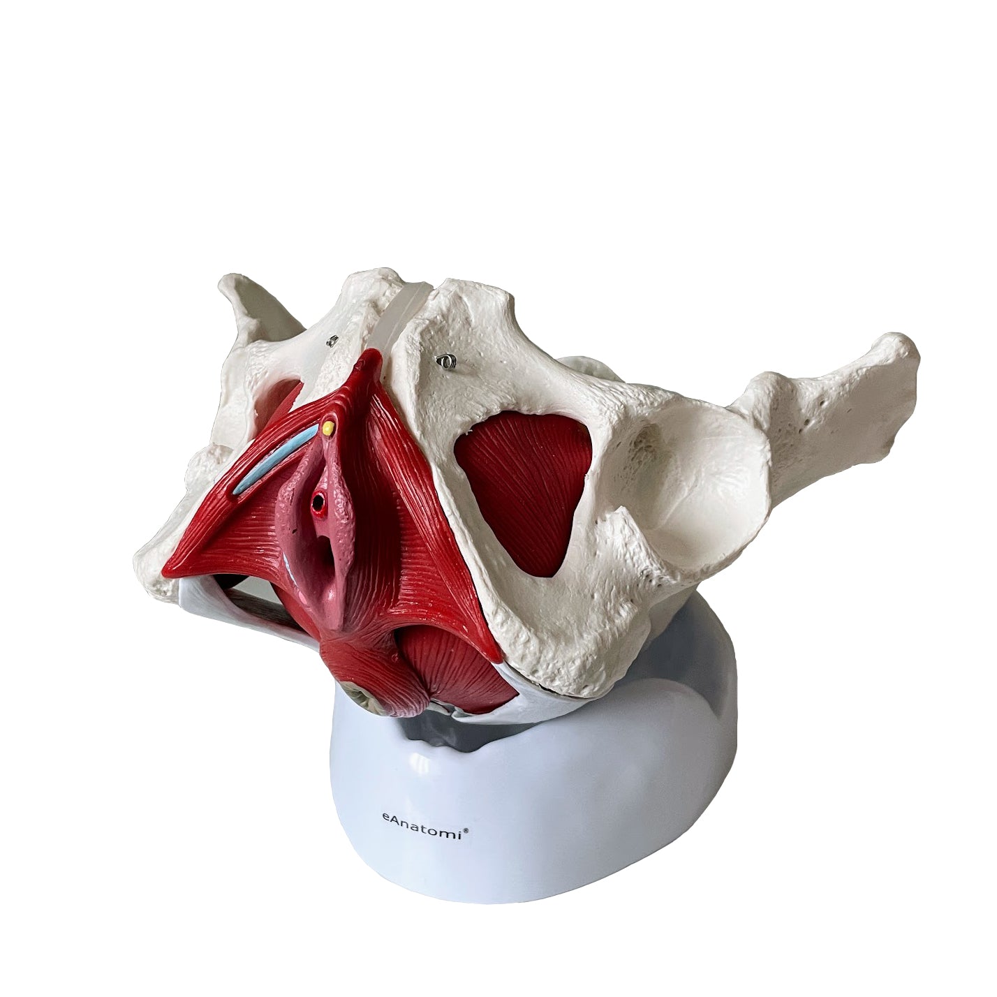
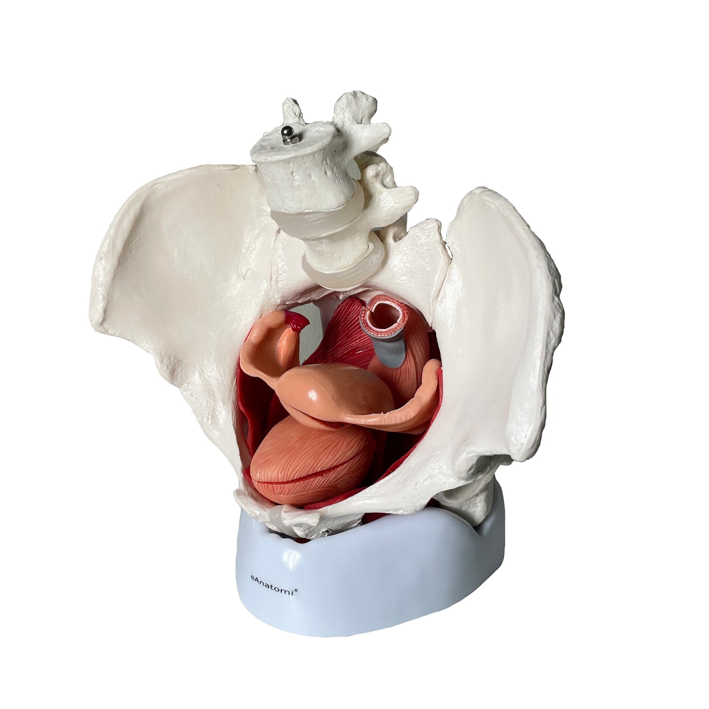
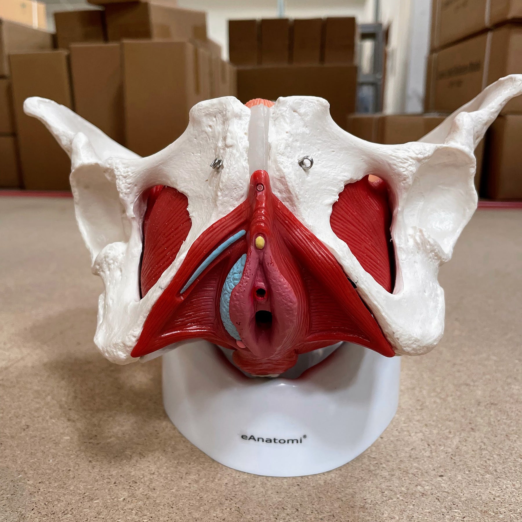
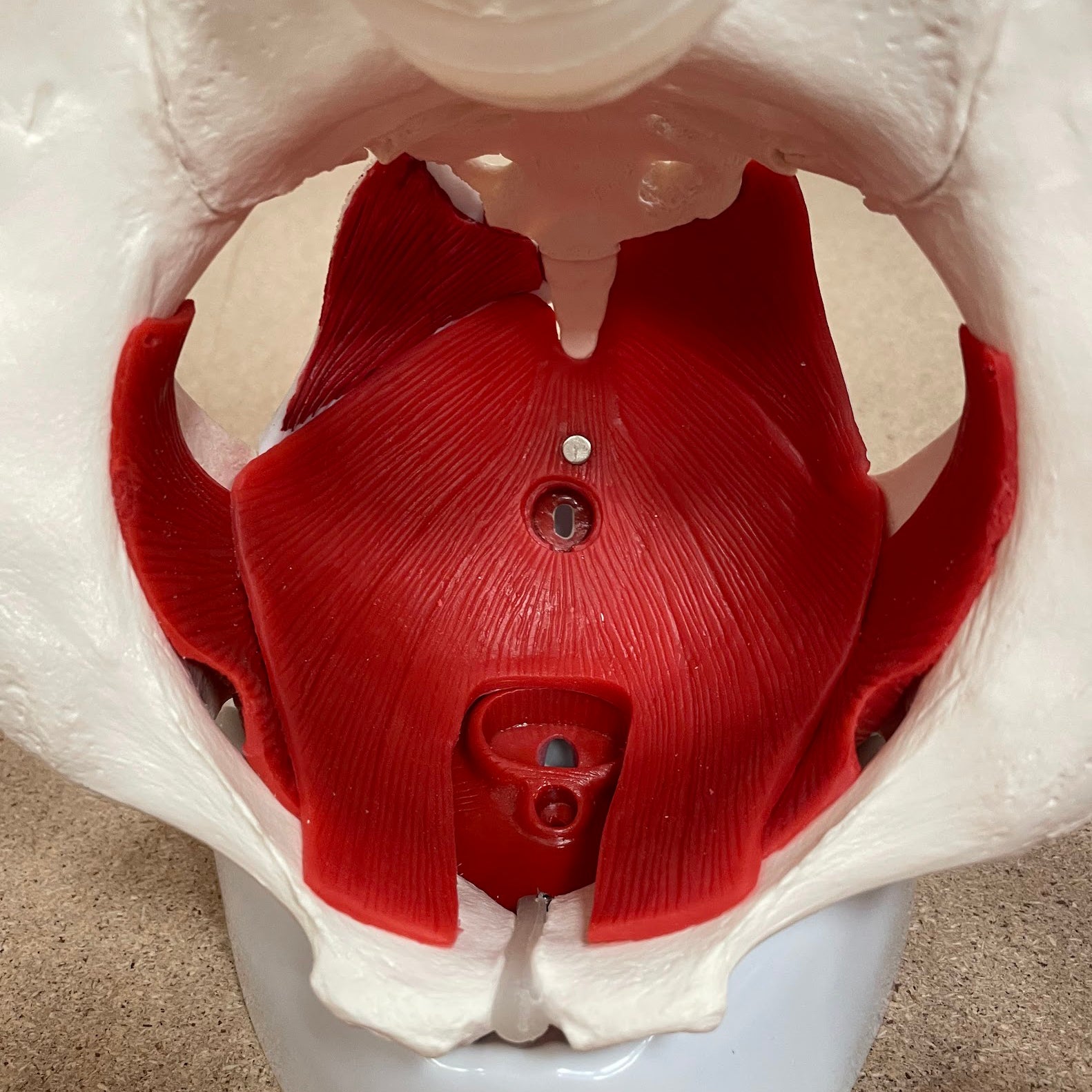
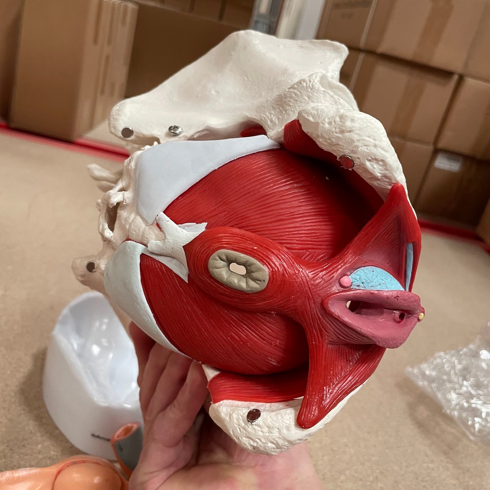
This cymbal model is particularly educational, practical and decorative. It shows the woman's pelvis with a focus on the pelvic floor and the internal and external genitalia.


The model is slightly reduced compared to an adult and corresponds in size to a 14-year-old. The pelvic ring, which is made up of bones, cannot be separated at all, although the sacrum can be opened. On the other hand, all the other organs can be taken out of the model and some of them can be separated because they are held together by discrete magnets (see the pictures on the left).


NB: As the model has been developed to be able to be separated in an easy and educational way as mentioned above. If you want for practical reasons that fewer parts can be easily removed, you can use a little glue to fix the parts that you do not need to remove - for example the ligaments on the back of the model.

Anatomically speaking

Anatomically speaking, the model shows both the woman's pelvis, the muscles in the pelvic floor, the internal and external genitalia as well as the relationships of the urinary bladder with the urethra and the rectum.


The pelvis consists of the 3 building blocks, which are the 2 hip bones and the sacrum. The model also shows the 2 lower lumbar vertebrae. Overall, the model shows the following bones:


Os coxae (the 2 hip bones) of which both are composed


- Os ilium (iliac bone/hip bone)

- Os ischii (seat bone)

- Os pubis (pubic bone)


Us sacrum (sacrum) incl. os coccygis (coccyx)
4th and 5th lumbar vertebra (lumbar vertebra)


In the bone tissue, the most important details can be seen, such as large nodules. Smaller details such as the linea glutaea are omitted. As something special, the sacrum can be opened. It makes it possible to see the canalis sacralis as well as the Y-shaped canals in its lateral wall (where nerves run). Furthermore, some ligaments are seen. It is first and foremost equal. sacrospinal and lig. sacrotuberal.


The pelvic floor (also called the pelvic floor or diaphragma pelvis in Latin) consists of the following muscles, which are clearly visible:


- M. levator ani (consisting of m. pubococcygeus and m. iliococcygeus)

- M. coccygeus


Other muscles are also seen such as the ischiocavernosus muscle, the bulbospongiosus muscle and the obturator internus muscle.


The internal genitalia include the ovaries (ovaries), the fallopian tubes (tubae uterinae), the uterus (uterus) and the vagina (vagina), all of which are seen in the greatest detail. Anatomically, it is located in the small pelvis. They are seen in relation to the urinary bladder (vesica urinaria) with the urethra (urethra) and the rectum (rectum).


The external genitalia are shown with the largest and most important details, which anatomically are located in front of and below the symphysis.


Many of these organs function as a channel or a reservoir (e.g. the vagina and the urinary bladder), which is why they are seen in a pedagogical way with an air-filled cavity (cavity). Special areas are shown in an educational color - for example glans clitoridis in yellow and fimbriae in dark red.

The pelvis as a birth canal


Normally the following 3 connection lines are specified:


- Conjugata vera (normally measures 11 cm in the female pelvis)

- Diameter obliqua (usually measures 12.5 cm in the woman's pelvis)

- Diameter transversa (usually measures 13 cm in the woman's pelvis)


On this pelvis model, all 3 above connecting lines measure 11 cm. This is due, among other things, to the fact that the size of the model corresponds to a 14-year-old (as mentioned at the beginning of the description above).

Movement-wise

In terms of movement, the pelvis is not flexible, which is why joints (e.g. the SI joints and the symphysis) cannot be separated or moved. The same applies to the joints in the 2 lower lumbar vertebrae.


When the internal organs of the model are separated, they are generally a bit flexible. For example, the urinary bladder can be squeezed quite a bit when you hold it. The pelvic floor, on the other hand, has a firmer consistency and is cast in one piece with the external genitalia and the rectal opening (can be seen in one of the pictures on the left).

Clinically speaking

Clinically speaking, the model can be used in connection with rehabilitation after childbirth, operative interventions, etc. It can also be used to understand diseases and disorders in the female genital organs, the urinary bladder and the rectum. Examples are urinary tract infection and tumors.

CUSTOMER SERVICE

custom-made items

If the product is stated as a made-to-order item, it means that the product is of such a size or such a high quality level that the product is only available on order. Delivery time may vary, but the price will always remain the same! Contact us for more information if you wish to order.

Prices and payment methods
  • Prices are stated in DKK including VAT.
  • You can pay with;
    - EAN no.
    - MobilePay
    - Visa, Mastercard
Right of withdrawal & return
  • The right of withdrawal is 14 days from delivery.
  • Return postage is 60,- when purchasing a label from us (sent as a PDF file via email) or 0,- when returning in person to our address.
  • See our terms and conditions here
Delivery / pickup

If the item is listed as in stock , it is physically located at our address in Frederiksberg and can therefore be shipped or picked up. When ordering, choose what you want.

If you wish to pick up the item, this can often take place immediately after ordering, but must be agreed with us. If you wish to have it shipped , this will be done via Postnord and often the same day the order is received.

If the item is listed as ' shipped directly from the factory ', the delivery time is usually 2-5 business days, but depends on the factory's stock status. If you want more precise information, you can always contact us before ordering.

Facts about eAnatomy
  • Founded in 2004 as a sole proprietorship and converted to ApS in 2019
  • 100% owned by Christian Birksø who is also responsible for the daily operations.
  • Develops and markets both original products, designed and produced by eAnatomi, and distributes many international brands.
  • Sells to both private and commercial customers in Denmark and abroad.

Other products selected especially for you

Our anatomical product range

Raffle for free products

All recipients of our newsletter are entered into raffles for free products. Sign up today and be in the running!