
Have you ever wondered how advanced and important your eye really is to your everyday life? This article gives you a detailed, easy-to-understand guide to how the eye is built and why it is relevant to know in 2026. When you understand the anatomy of the eye, it becomes easier to understand vision, prevent eye diseases and keep up with new technology. We delve into the main parts of the eye, their functions, the process of vision itself, development throughout life and future research. Read on and learn more about the fascinating world of the eye.
The Main Parts of the Eye and Their Functions
The eye is one of the most complex organs in the body, and to understand how the eye is built, we need to take a closer look at each part and its function. Each structure works closely together to give us clear vision and protect the eye from damage. In this section, you will get a detailed overview of the most important outer and inner parts, the retina and the optic nerve, and the interaction between them. This gives you a solid foundation for understanding how the eye is built and why this knowledge is central to both health and technology.
External structures of the eye
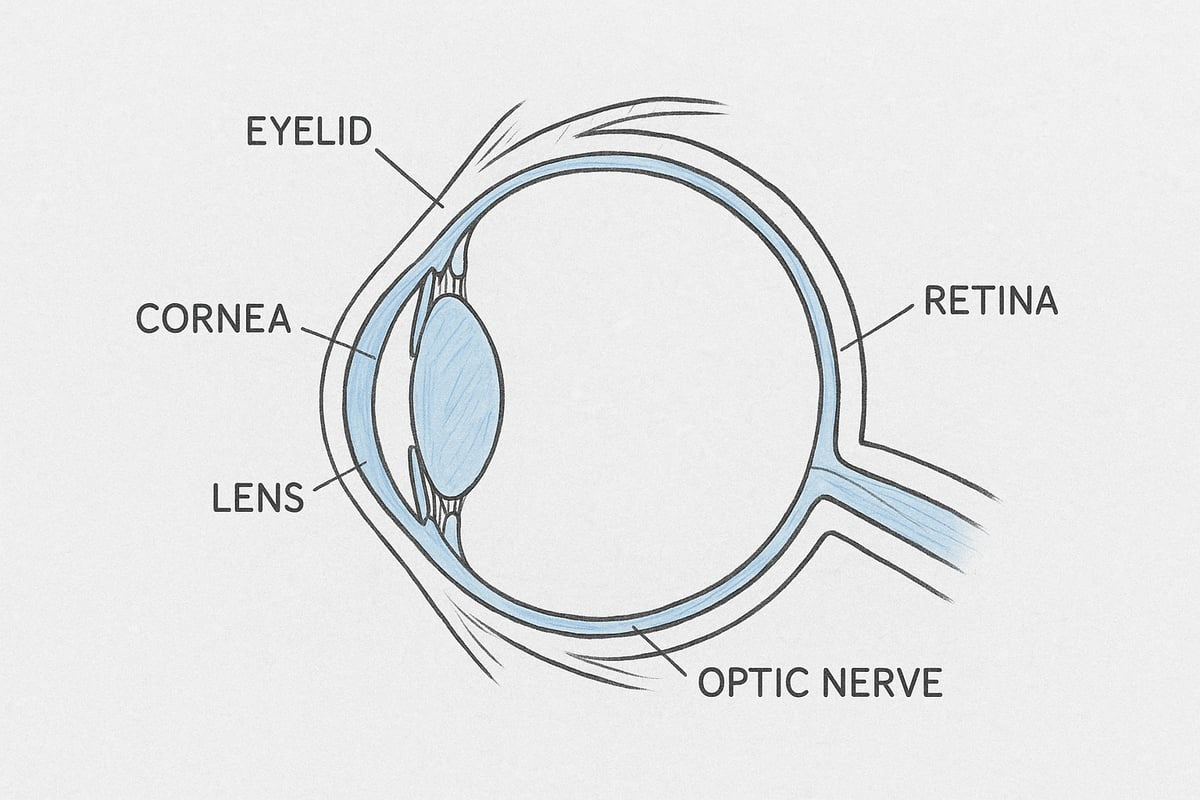
When we look at the structure of the eye, we start with the external structures that protect and maintain the health of the eye. The eyelid acts as a barrier that protects the eye from dust and foreign objects. Together with the eyelashes, it helps prevent particles from hitting the surface of the eye.
The conjunctiva, also known as the white of the eye and the inside of the eyelid, covers the white of the eye and the inside of the eyelid. This thin membrane protects against infection and keeps the eye moist. The lacrimal gland produces tears, which cleans and lubricates the eye, which is essential for comfort and quality of vision.
The sclera, the white of the eye, provides support and protection for the internal structures. The cornea is the clear front of the eye, which acts as a powerful lens and refracts light so that it can be focused further into the eye.
Common problems in the external structures include dry eyes and conjunctivitis (eye inflammation). According to Danish health data, approximately 20 percent of adults experience dry eyes, which can cause discomfort and blurred vision.
To visualize and understand these external structures, you can examine anatomical models of the eye , where the individual parts are clearly marked and explained.
Internal structures of the eye
To truly understand how the eye is constructed, we need to look at the internal parts that focus and regulate light. The lens sits behind the pupil and changes shape to focus light precisely on the retina. This ability to adapt is essential for sharp vision at different distances.
The iris is the colored part of the eye that controls how much light comes in by changing the size of the pupil. The ciliary body is the muscle that adjusts the shape of the lens and produces aqueous humor, which keeps the pressure in the eye stable.
The vitreous is a jelly-like mass that fills most of the eye and helps maintain the eye's shape. The aqueous humor circulates between the anterior and posterior chambers and is important for nutrition and pressure regulation.
If any of these structures do not function properly, it can lead to diseases such as cataracts, where the lens becomes cloudy, or glaucoma, where pressure in the eye increases and damages the optic nerve. Modern treatments, especially for cataracts, have made it possible for many to regain their vision after surgery.
The retina and optic nerve
When we examine how the eye is structured, the retina and optic nerve play a central role in the visual process itself. The retina consists of several layers, the most important of which are the photoreceptors: rods and cones. Rods detect light and darkness, while cones provide color vision and detail vision.
The optic nerve connects the eye to the brain and transmits the electrical signals that arise in the retina to the visual center. In the center of the retina is the yellow spot (macula), which is responsible for the sharp, central vision we use to read and recognize faces.
One of the most common diseases of the retina is age-related macular degeneration. This condition is the most common cause of vision loss in people over 65 in Denmark and can lead to severe problems seeing details.
The interaction between the parts of the eye
To understand how the eye is constructed, we also need to look at how all these parts work together. Light travels through the cornea, pupil, lens, and vitreous humor before reaching the retina. Each part has a specific role, and their precise cooperation is necessary for clear vision.
The optical axis is the line that light follows through the eye. If the cooperation between the structures does not function optimally, problems such as strabismus or astigmatism can arise, where light is not focused correctly on the retina. This can cause double vision or blurred vision, and shows how vulnerable the system is to small imbalances.
How Vision Works: From Light to Image
Vision is the result of a close interaction between the anatomy of the eye and the brain's ability to interpret impressions. But how is the eye structured so that we can experience the world in colors, shapes, and movement? To understand the miracle of vision, we must follow the journey of light from when it hits the eye until it is transformed into images in the brain.
Steps of the vision process
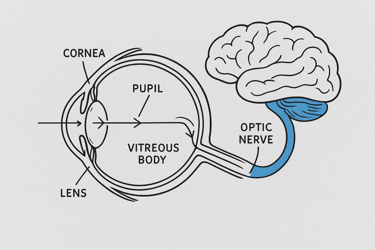
When we ask how the eye is constructed, it is natural to start with the journey of light. First, the light passes through the cornea, which refracts and focuses the rays. Then, the light passes through the pupil, the size of which is controlled by the iris and adapts to the amount of light.
Behind the pupil is the lens, which further adjusts focus so that sharp images land on the retina. The vitreous humor helps guide light and supports the shape of the eye. The retina contains millions of photoreceptors, rods and cones, which convert light into electrical signals.
These signals are sent through the optic nerve. The brain receives and processes the signals so that we experience an image. A classic example of how the eye is structured and works is seen in the use of glasses: In the case of nearsightedness or farsightedness, glasses correct refractive errors so that the light hits the retina correctly.
Color vision and night blindness also depend on the function of photoreceptors. The cones detect colors, while the rods are sensitive to light and allow us to see in the dark. If the balance between these cells is disturbed, it can lead to reduced night vision or color blindness.
The brain and visual perception
The structure of the eye is not only determined by the parts of the eye, but also by its interaction with the brain. When signals from the retina reach the optic nerve, they are sent to the primary visual cortex in the occipital lobe. This is where the information is collected and interpreted so that we understand what we see.
The brain can create optical illusions and deceptions because it tries to fill in missing information or reinterpret impressions. Over 30% of the cerebral cortex is involved in visual processes, underscoring the importance of vision to our experience of the world.
It's fascinating how new technology can expand our understanding of how the eye is built. If you want to see how the brain and vision are connected, you can find anatomical posters about the brain and nervous system that visualize this complex process.
Errors in the visual process
When we look more closely at how the eye is constructed, it becomes clear that small errors can significantly affect vision. Myopia (nearsightedness) is caused by the eyeball being too long, so the image is formed in front of the retina. Hyperopia (farsightedness) occurs when the eyeball is too short, and the image is formed behind the retina. Astigmatism is caused by irregularities in the cornea or lens.
Symptoms of refractive errors include blurred vision, headaches and fatigue. Correction options range from glasses and contact lenses to modern laser surgery. It is noteworthy that the incidence of myopia among young people is increasing globally, which emphasizes the importance of knowledge about how the eye is built.
Development and Changes in the Eye Throughout Life
The eye is constantly changing throughout life. Regardless of how the eye is structured, significant changes occur from childhood to old age, affecting both vision and eye health. By understanding these developmental stages, we can better protect vision and detect problems early.
Eye development in children
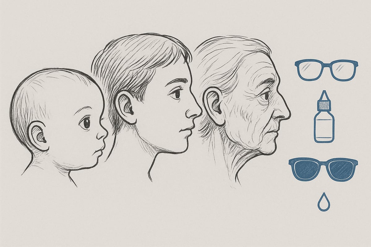
In infants, vision is blurred at birth, but develops rapidly in the first few years of life. Already during the first months, the anatomy and function of the eye becomes more complex, and the child begins to recognize colors and shapes. Parents often ask how the eye is built in young children, and the answer is that the important structures gradually mature, so that visual acuity improves.
It is crucial to have early eye exams, as problems such as strabismus or amblyopia (lazy eye) can develop without any visible symptoms. If detected early, treatment can give the child optimal vision development. Statistics show that approximately 5% of Danish children have vision problems when they start school. Regular check-ups can ensure that the child gets the best start in life with clear vision.
Age-related changes
As we age, the eye's ability to focus changes. Many people experience presbyopia, where it becomes more difficult to see clearly up close, typically from their mid-40s. This is due to the way the eye is built, especially the lens and muscles, which lose flexibility. The result is the need for reading glasses or other aids.
Cataracts also become more common with age. The lens becomes cloudy, affecting the quality of vision. Macular degeneration, which affects the central part of the retina, can lead to a loss of detailed vision. These changes can cause challenges in everyday life, such as reading or recognizing faces. However, early detection and treatment can make a big difference.
Eye diseases and prevention
Several diseases can affect the eye throughout life. The most common are glaucoma, cataracts and diabetic retinopathy. Many people ask how the eye is structured when diseases occur, and the answer is that small changes in pressure, blood vessels or lens can lead to vision loss.
Prevention is essential. Good advice includes wearing sunglasses, a healthy diet rich in vitamins, and regular eye exams. Research and new treatment methods are constantly being developed, and a Danish research project on chronic eye diseases is working on better treatment and prevention. Taking responsibility for your eye health can reduce the risk of serious problems.
Environmental impacts and lifestyle
Our lifestyle significantly affects eye health. Today, many people spend time in front of screens, which can cause digital eye strain. Over half of Danes experience tired eyes after long screen time. How is the eye structured when exposed to such influences? The eye muscles and tear glands are strained, which can lead to dryness and discomfort.
Smoking increases the risk of serious diseases such as cataracts and macular degeneration. A diet rich in vitamin A and omega-3 fatty acids strengthens the cells of the eye and protects against damage. Small changes in everyday life can therefore have a major impact on vision and eye health throughout life.
Anatomy of the Eye in Research and Technology 2026
Research into the structure of the eye has advanced significantly in recent years. Modern technology and scientific discoveries have opened new doors for understanding, treating, and teaching the anatomy of the eye. Here you will gain insight into the most groundbreaking trends shaping the future of eye health.
Latest scientific discoveries
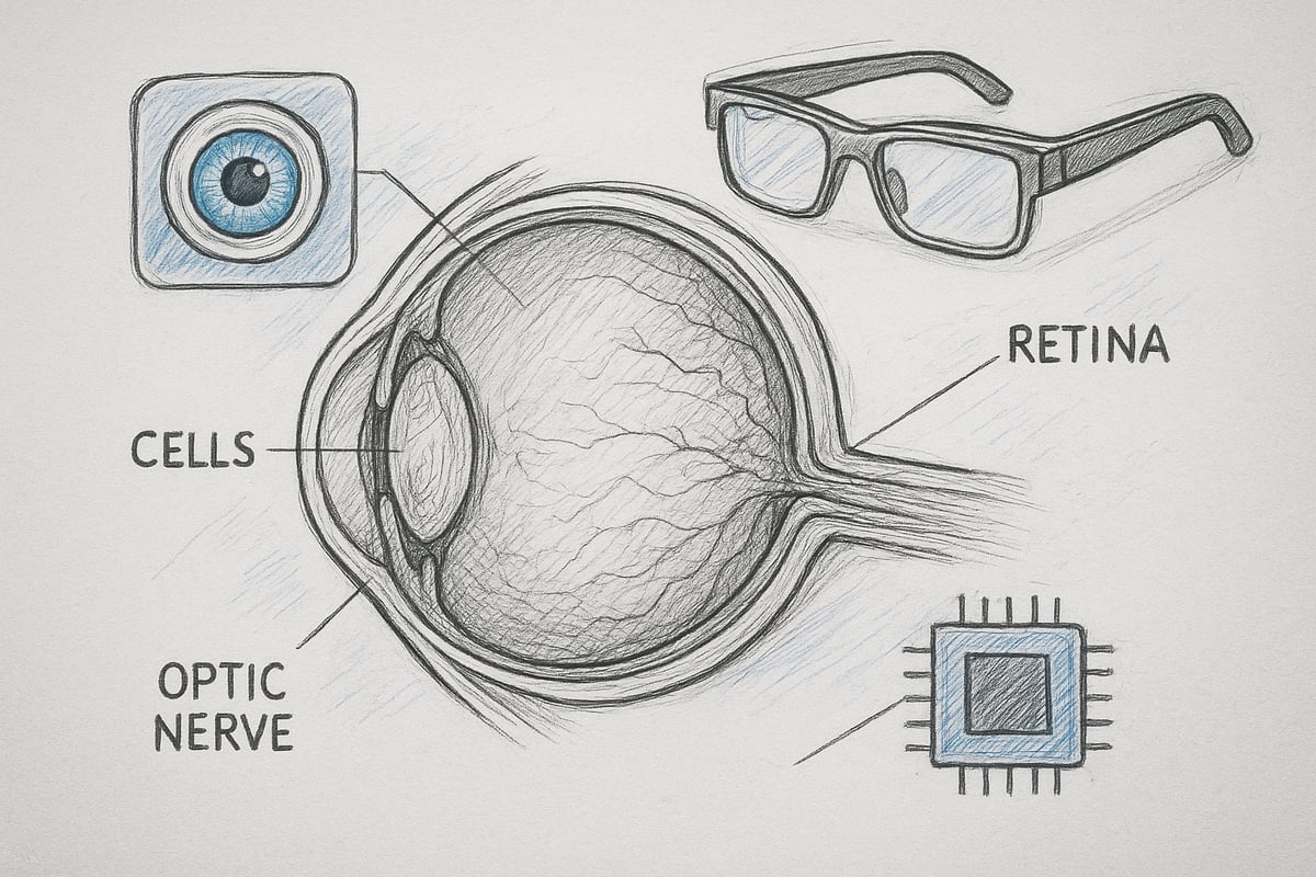
In 2026, researchers are deeply concerned with mapping out how the eye is structured down to the molecular level. New techniques make it possible to analyze the eye's cells and their interactions, providing insight into how vision is formed and maintained.
Genetic engineering has revolutionized the treatment of inherited eye diseases. CRISPR trials for retinitis pigmentosa have brought hope to patients who previously had no treatment options. Scientists are working to understand how mutations in specific genes affect the structure and function of the eye.
The number of published studies on how the eye is built has increased by 25% since 2020. This growth is evidence of an intensified global effort to uncover the secrets of the eye and develop new treatment methods.
Innovative eye surgery and treatment
Advances in eye surgery have changed how the structure of the eye can be improved and reconstructed. Laser surgery and advanced lens implants are now routinely used to correct refractive errors and cataracts.
Artificial retinas and bionic eyes have undergone major advances. In 2023, the first Danish patient received a bionic eye, marking a breakthrough for vision restoration. These technologies are based on the understanding of how the eye is structured and how electrical signals can be transmitted to the optic nerve.
The future offers new opportunities where biological and technological solutions merge. Research focuses on creating even more precise and gentle treatments that can be tailored to the needs of the individual patient.
Digital aids and vision technology
Digital developments have also changed how the structure of the eye is understood and handled in everyday life. Smart glasses and augmented reality give the visually impaired access to information and navigation that was previously unthinkable.
Vision training and eye health monitoring apps have become popular. They can alert the user to early signs of disease, improving prevention options. A good example is EyeAI: AI-assisted eye disease detection , where artificial intelligence analyzes images of the eye to identify potential problems quickly and accurately.
Statistics show that 1 in 10 Danes now uses digital vision aids. This development highlights how closely technology and knowledge about the structure of the eye are connected in modern vision care.
Anatomy of the eye in education and communication
Education about the structure of the eye has also been given a boost with new visual aids. Detailed models and posters make it easier for both students and patients to understand the complex structure of the eye.
3D visualizations and virtual eye models are used especially in medical education and patient education. They make it possible to explore the inside of the eye and see how diseases affect the individual parts. Many educators use anatomical posters about sensory organs to illustrate the process of vision and the main parts of the eye.
This communication is essential to raising awareness about eye health and emphasizes the importance of knowing the answer to the question: how is the eye structured?
Frequently Asked Questions about the Structure of the Eye
When we examine how the eye is structured, many questions often arise. Here are the most common answers, so you get a solid overview of the eye's anatomy, function, and health.
What parts does the eye consist of and what are their functions?
To understand how the eye is built, you should know the most important parts. The eye consists of external and internal structures that work closely together. The external parts such as the eyelids, eyelashes and conjunctiva protect the eye from dust and bacteria. Inside we find the cornea, lens and retina, which focus light and convert it into images.
The retina contains photoreceptors that detect light and color. The optic nerve transmits information to the brain. If any of these parts are damaged, vision can be significantly affected. For a comprehensive overview of sensory organs, please read this overview of sensory organs .
How does the eye protect itself?
When asking how the eye is constructed, it is important to focus on the eye's natural protective mechanisms. The eyelids close automatically in case of danger, shielding the eye from foreign objects. The eyelashes catch dust particles before they reach the surface of the eye.
The tear glands produce tears that keep the eye moist and wash away small particles. The blink reflex is triggered very quickly if something approaches the eye. These functions ensure that the eye remains healthy and resistant to infection and irritation.
What happens when you become nearsighted or farsighted?
Many people ask how the eye is structured when they experience vision changes such as nearsightedness or farsightedness. In nearsightedness (myopia), the eyeball is often too long, so the image is formed in front of the retina, making it difficult to see things at a distance. In farsightedness (hyperopia), the eye is too short, so the image is formed behind the retina, making it especially difficult to see things up close.
These conditions are called refractive errors and can be corrected with glasses, contact lenses, or laser surgery. Properly adjusted eyeglass prescription significantly improves vision and restores balance to the eye's optical system.
How does the eye change with age?
As we age, the structure of the eye gradually changes. A common change is presbyopia, where the lens loses its elasticity, making it harder to focus up close. Many people start needing reading glasses around the age of 40-45.
The risk of cataracts and age-related macular degeneration also increases with age. These changes can affect everyday vision, but early detection and treatment can often help preserve vision for as long as possible.
What diseases most often affect the eye?
When exploring how the eye is built, it is relevant to know the most common eye diseases. The most common are cataracts, glaucoma and age-related macular degeneration. Symptoms can include blurred vision, visual field loss or eye pain.
| Disease | Typical symptoms | Prevention |
|---|---|---|
| Cataracts | Blurred vision, glare | Sunglasses, check |
| Glaucoma | Visual field loss, pressure | Regular measurement |
| Macular degeneration | Loss of detail vision | Healthy eating, check |
Regular eye exams and healthy habits can reduce the risk of many eye diseases.
Can you strengthen or protect your eyesight?
Some people ask how the eye is built and whether there is anything you can do to improve your vision. The answer is yes. A healthy diet with vitamin A and omega-3 fatty acids is important for eye health. Limit screen time and take regular breaks to avoid eye strain.
Wear sunglasses with UV protection and get your eyes checked regularly. Regular exams can detect problems early and prevent permanent damage. Small changes in your daily routine can make a big difference to your vision.
Now that you have gained insight into the fascinating structure of the eye, its lifelong development and the latest research, it is natural to be curious about how you can learn even more yourself or convey knowledge about the eye to others. At eAnatomi you will find detailed anatomical models and posters that make it easy to visualize and understand the complex structures of the eye — whether you are a student, teacher or just interested in health. If you want to dive deeper and discover more innovative teaching tools, you can Read more here
0 comments