

Imagine a time when our knowledge of the body was characterized by myths and misunderstandings, and where medical science groped in the dark without precise tools.
With a historical article about the use of anatomical models, we open the door to a world where curiosity and innovation have transformed teaching and research.
In this article, you will gain insight into the fascinating origins of models, the development of materials, their role in education, technological advances, ethical dilemmas, and future possibilities.
Be inspired to explore how anatomical models have paved the way for understanding, learning, and medical advancement.
The origin of anatomical models
Imagine a time when knowledge of the inside of the body was shrouded in taboo and mystery. In ancient Greece and Egypt, understanding of anatomy was limited, often influenced by religious beliefs. Many cultures forbade the dissection of human bodies, making it almost impossible to learn about the structure of the body. This challenge haunted scholars and laypeople alike for centuries, and it was not until the Renaissance that anatomy began to develop in earnest.
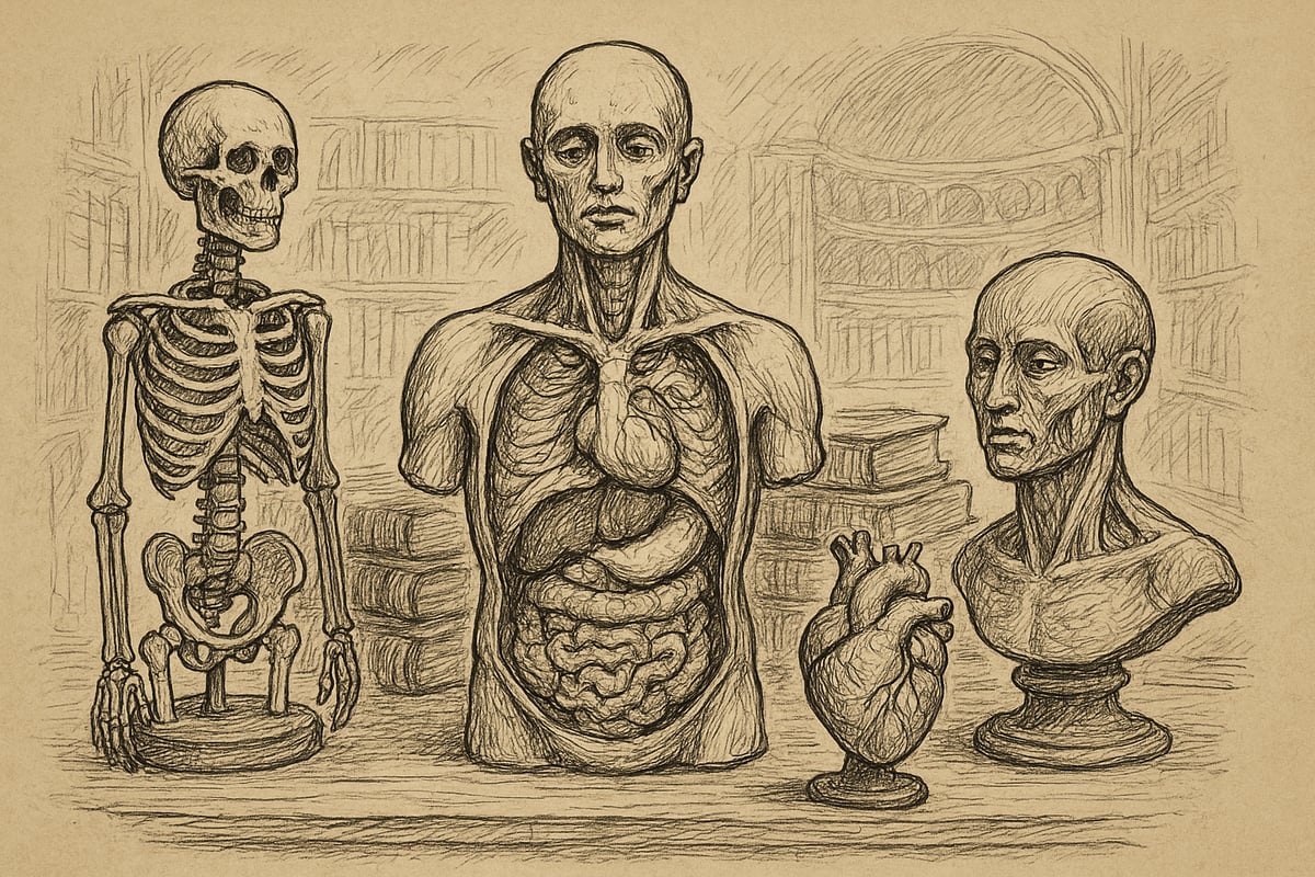
During this period, the need for alternative teaching aids arose. The first anatomical models were created in Europe and Asia in response to the restrictions placed on access to cadavers by religious and cultural norms. Models were made from materials such as wax, wood, and clay. These materials were available and easy to shape, making it possible to reproduce the structures of the body in an educational way. Already here, the basis for a historical article on the use of anatomical models began to take shape.
Wax models became particularly popular in Italy in the 17th and 18th centuries. Famous collections such as La Specola in Florence still contain outstanding examples of this art form. Wax had the advantage of being able to reproduce fine details and colors, which made the models both realistic and educationally valuable. Statistics from the period show that the limited access to cadavers in the 16th-18th centuries led to a significant increase in the use of anatomical models. This helped to make a historical article on the use of anatomical models extremely relevant for understanding the development of medical education.
The use of models was not only a matter of circumventing ethical and religious barriers. They also served as important tools in public education. Anatomical models were exhibited in museums and anatomical theaters, where both students and citizens could gain insight into the mysteries of the body. For many, it was the first time they had the opportunity to see the internal structure of the body. This pedagogical approach has been central to a historical article on the use of anatomical models, because it illustrates how the models paved the way for new understanding and wider acceptance of medical knowledge.
The importance of models for medical education cannot be overstated. They made it possible for students to learn anatomy without access to real cadavers, and they helped to standardize teaching across national borders. Today you can read more about the history of anatomical models , where you will get an overview of the development and the many types of models that have characterized teaching throughout time. A historical article on the use of anatomical models thus shows how these unique tools have shaped both research, education and society's view of anatomy.
Development of materials and techniques
The development of materials and techniques has been crucial to how a historical article on the use of anatomical models can be understood. The models have changed significantly from the first, primitive versions to today's advanced solutions. Over time, the choice of materials and technological advances have set the standard for both level of detail, durability and pedagogical utility.
From wax to modern synthetic materials
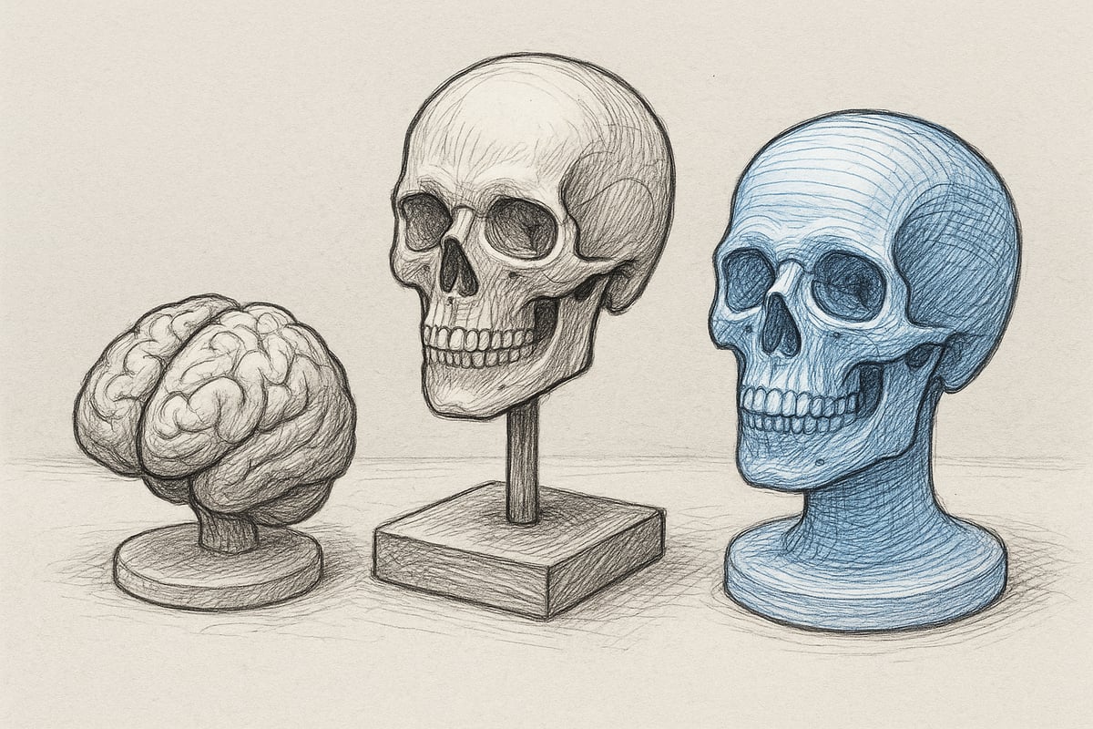
In a historical article on the use of anatomical models, it is clear how the choice of materials has reflected the technological level of the time. The first models were often made of wax, as this material could reproduce soft tissues and fine details in a way that was unattainable with stone or wood. Wax models became especially popular in the 17th and 18th centuries, when museums, such as La Specola in Florence, created lifelike reproductions of the human body's interior.
However, wax had its limitations. The material was sensitive to temperature and could melt or deform, making storage and transportation challenging. To meet the need for more robust models, wood and papier-mâché began to be used in the 18th and 19th centuries. Wooden models were more durable and suited to repeated use in teaching, while papier-mâché allowed for cheaper mass production.
With the industrial development of the 20th century, plastic and silicone were introduced. These materials revolutionized the field, combining flexibility with durability and allowing the production of anatomical models on a large scale. A historical article on the use of anatomical models will often highlight how plastic models set new standards for detail and hygiene, as they could also be washed and disinfected.
Today, 3D printed models have become an integral part of education, increasing accessibility and individualizing the learning experience. Statistics show that the production of synthetic models increased significantly from the 1960s onwards, which made it possible for more educational institutions to integrate them into their curricula. A historical article on the use of anatomical models thus illustrates how the development of materials has affected the quality of education and pedagogical value.
To compare the properties of the materials, the following table can be used:
| Material | Durability | Level of detail | Price | Usage history |
|---|---|---|---|---|
| Wax | Low | High | High | 17th-19th centuries |
| Wood | High | Medium | Medium | 18th-19th century |
| Papier-mâché | Medium | Low | Low | 19th-19th century |
| Plastic | High | High | Low | 1960s and onwards |
| Silicone | High | Very high | Medium | 1980s and onwards |
| 3D printing | Very high | Very high | Varied | 2000s and beyond |
The development in materials illustrates how a historical article on the use of anatomical models encompasses both technological advances and changing pedagogical needs.
Technological breakthroughs and innovations
Technological breakthroughs have helped transform how a historical article on the use of anatomical models can document the development of the field. The introduction of 3D scanning and printing has made it possible to produce precise copies of individual anatomical structures, giving students and healthcare professionals access to unique learning tools.
Today, the integration of AR and VR technologies has become a central part of modern anatomy education. Digital models and virtual simulations allow for interactive learning, where the user can examine the body layer by layer or practice surgical skills without risk. According to a competitor analysis, 70% of medical faculties in Europe now use digital models to supplement traditional solutions. This shift reflects a growing recognition that technology can enhance understanding of complex anatomical relationships.
Examples of these innovations include the use of hybrid models, where physical and digital elements are combined to create a more nuanced learning experience. Personalized models based on patient data are also gaining ground, especially in the context of surgical planning and specialized training. To illustrate this development, read more about The Evolution of 3D Dissection Models , where modern technologies demonstrate the latest possibilities in the field.
Looking ahead, there is no doubt that a historical article on the use of anatomical models will have to deal with an increasingly digitalized reality. With hybrid solutions and personalization, the models will not only become more accessible, but also more relevant to the individual user. Developments indicate that pedagogical innovation and technological progress will go hand in hand in the coming years.
The role of anatomical models in teaching and research
Anatomical models have played a crucial role in bridging the gap between theory and practice in medical education. In a historical article on the use of anatomical models, it is clear that models have both enhanced learning and created new opportunities for research and continuing education.

Importance for medical education
The use of anatomical models has always been central to medical education. From the earliest days, when access to cadavers was limited, models were developed to illustrate the structures of the body. In a historical article on the use of anatomical models, we see how these tools became indispensable, especially in universities and hospitals.
Over time, models have served as both a supplement and an alternative to cadaver dissection. Students can repeat exercises, investigate complex anatomical relationships, and gain understanding without relying on rare or ethically challenging cadavers. This makes models ethically sound and practically applicable tools.
A concrete example is the use of anatomy models for medical students , where both basic and advanced structures can be studied in detail. According to a 2023 survey, 85% of medical students consider models to be essential to their learning process. This statistic supports why a historical article on the use of anatomical models remains relevant today.
Models contribute not only to basic understanding but also to specialized continuing education. Clinical skills laboratories and simulation training use models to prepare students and professionals for real-life situations. Countries and education systems have different traditions, but the pedagogical value of models is internationally recognized.
Below is an overview of areas of application:
| Application area | Benefits |
|---|---|
| Universities | Repeatability, details |
| Hospitals | Training, continuing education |
| Clinical laboratories | Simulation, specialization |
In a historical article on the use of anatomical models, it is clear that these tools have strengthened both knowledge and practice.
Development of pedagogical methods
The development of pedagogical methods has followed the same pace as the technology behind the models. Originally, teaching was often characterized by passive observation, where students watched while the teacher demonstrated on a model. Over time, the focus has shifted towards active learning.
A historical article on the use of anatomical models highlights that hands-on and interactive exercises significantly enhance learning. Simulation training and the use of skill trainers have been shown to reduce errors in practice. Statistics show that surgical errors can be reduced by up to 60% after simulation training. This emphasizes the effectiveness of models in both basic and advanced training.
Models allow students to work independently and repeatedly, strengthening both visual memory and comprehension. In a historical article on the use of anatomical models, we see how this approach has changed the way medicine and health sciences are taught.
In summary, models have not only improved knowledge acquisition, but also made it possible to adapt teaching to different learning styles and needs. The continued development of pedagogy will undoubtedly maintain the central role of models.
Ethical, cultural and societal aspects
Throughout history, ethical issues surrounding the dissection of human cadavers have been central to a historical article on the use of anatomical models. Many societies have had moral, religious, and legal reasons for restricting access to dead bodies. This led doctors and researchers to seek alternatives, which had a major impact on the development of anatomical models.
During the Renaissance, cadaver dissection was challenged by religious dogma and societal norms. One of the most prominent figures, Andreas Vesalius, paved the way for modern anatomy by insisting on empirical studies, but he faced considerable resistance. You can read more about Andreas Vesalius' contributions to anatomy , which illustrates how ethical dilemmas have been crucial to innovation. For many, wax models and later other materials were ethically sound alternatives, making it possible to convey anatomical knowledge without violating religious or cultural boundaries.
Cultural and religious differences have had a major impact on how anatomy was taught and understood. In some countries, strict religious prohibitions against touching or dissecting dead bodies have meant that educators have had to find creative solutions. Statistics show that up to 90% of schools in some regions use anatomical models exclusively for ethical reasons. In a historical article on the use of anatomical models, we see how this has led to a wider acceptance and development of pedagogical tools that respect local norms.
The societal consequences of model use extend far beyond teaching itself. Anatomical models have made knowledge more accessible and inclusive, as they do not require access to human cadavers, which promotes equality in learning situations. In addition, models have helped to educate the general public, not just professionals. For example , anatomical posters in education have played a significant role in conveying complex knowledge in an easily understandable way. In a historical article on the use of anatomical models, it becomes clear that models contribute to breaking down barriers, increasing diversity, and creating societal acceptance of medical knowledge.
Modern applications and future possibilities
Today we are in the midst of a revolution, where a historical article on the use of anatomical models is more relevant than ever. Developments in technology and pedagogy have brought models to the center of both teaching, research, and patient communication. This section highlights how modern applications and future possibilities are shaping our understanding of anatomy and medical practice.
Integration into health education and patient communication
A historical article on the use of anatomical models clearly illustrates how models have become indispensable in modern healthcare education. Today, they are used not only in medical school, but also in dental and veterinary education, as well as in hospitals and clinical skills laboratories.
Students are given the opportunity to explore complex structures through both physical and digital models. This increases understanding and facilitates the transition from theory to practice. Many educators use models to demonstrate disease processes, anatomical variations, and treatment options.
Patient communication has also changed significantly. Doctors often use models to explain diagnoses and treatment plans. Statistics show that 75% of doctors find that patients understand their situation better when models are included in the conversation.
Integrating anatomical models into teaching and patient communication results in more inclusive and effective learning. A historical article on the use of anatomical models documents this development and its positive effect on both students and patients.
Technological trends and innovations
Technological advances have transformed the possibilities for learning and training. A historical article on the use of anatomical models highlights how 3D printing, virtual reality, and interactive digital models are now standard in many institutions.
For example, advanced 3D printers such as the Stratasys J850 Digital Anatomy 3D Printer are used to create realistic and personalized anatomical models. These models make it possible to simulate surgical procedures and plan complex operations with high precision.
Virtual reality and AR technologies allow students to explore the body in new ways. The interactive 3D head model for medical devices is an example of how digitization can make anatomy education more vivid and accessible.
The market for 3D printed anatomical models is growing by 15% annually, reflecting the increasing demand for innovative solutions. A historical article on the use of anatomical models shows how technology has opened the door to new pedagogical methods and effective patient treatment.
Future perspective: From the classroom to the digital world
Looking ahead, it becomes clear that a historical article on the use of anatomical models can only hint at the full potential. Developments point towards even more advanced hybrid models that combine physical materials with digital layers and personalized solutions based on patient data.
The future of anatomy education will likely take place in both physical and virtual environments. Simulation training with customized models will improve skills and reduce errors. Furthermore, patient communication will become even more visual and understandable with the help of interactive models.
An important aspect is the ethics behind model development. Focus on sustainability, inclusion and access for all user groups will characterize both research and practice. A historical article on the use of anatomical models will in the future be able to document how these tools have contributed to equality, innovation and global knowledge sharing.
When we look back at the exciting development of anatomical models, it becomes clear how important they have been for both teaching and research over time. You and I can today benefit from the many technological advances that have made the models more realistic, accessible and safe for all user groups. If you are curious to explore how modern models can make a difference in your work or study, you can delve even deeper into the possibilities, innovations and wide selection at eAnatomi ApS.
Read more here
0 comments