

The brain remains one of the body's most complex organs, and in 2025, understanding its details is more important than ever. An anatomical brain model can make even the most advanced structures tangible for students, educators, healthcare professionals, and curious individuals alike.
This guide will help you choose, understand and use the right model. We will review everything from definition and types to application, selection criteria, maintenance and future possibilities. Get a handle on the anatomy of the brain and strengthen your knowledge in practice.
What is an anatomical brain model?
Definition and purpose
An anatomical brain model is a three-dimensional teaching tool that allows the brain's complex structure to be visualized in physical form. The model differs from digital images and posters by providing the user with a concrete and tactile experience. With an anatomical brain model, both students and healthcare professionals can examine the structure of the brain, identify specific areas, and understand functional relationships.
The purpose is to make anatomy more tangible and easy to understand. The model is used in teaching, patient guidance and research, where it helps illustrate everything from anatomical details to disease progression. You can find a wide selection of models at Brain Models and Anatomical Collections , where different types and sizes are available.
Historical development and technological progress
The development of anatomical brain models began with simple wax models used in medical schools over 100 years ago. Later, plastic, silicone, and resin became popular materials, giving the models increased durability and detail. Today, advanced 3D printing techniques are used to allow the models to be customized to specific teaching needs and to show even the smallest structures.
Statistics show that Danish educational institutions and hospitals are increasingly investing in modern brain models. This is due to both technological advances and an increased focus on interactive learning.
Advantages of physical versus digital model
Physical models and digital 3D models each have their strengths, but also limitations. A physical anatomical brain model offers tactile feedback, which strengthens spatial understanding and makes it easier to remember complex relationships. Digital models offer flexibility and the ability to zoom in and rotate structures, but lack the physical interaction.
| Property | Physical model | Digital model |
|---|---|---|
| Tactile learning | Yes | No |
| Mobility | Limited | High |
| Level of detail | High | Very loud |
| Price | Often higher | Often lower |
For many, the combination of both types provides the best learning results.
Key anatomical details
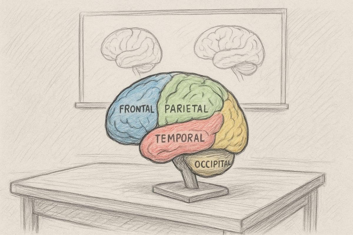
A typical anatomical brain model represents the main parts of the brain: the cerebrum, the cerebellum, and the brainstem. The models often include color coding and clear labels so that lobes, blood vessels, and nerves can be easily identified. Several models can be disassembled into segments, allowing for the study of internal structures and connections.
- Color-coded lobes: Frontal, parietal, occipital, temporal
- Removable parts: Brain stem, cerebellum
- Visible blood vessels and nerves
This structure ensures that even complex details are clear.
Target groups for brain models
An anatomical brain model is used by several different user groups. Students of medicine, physiotherapy and biology use models in teaching to enhance understanding. Teachers use them to convey complex topics visually and interactively. Healthcare professionals use models for patient guidance, where they can demonstrate the course of a disease or operations.
Individuals with an interest in anatomy are also choosing to invest in an anatomical brain model. Statistics show a significant increase in demand among medical students and physiotherapists, which underlines the model's importance as a learning tool.
Types of anatomical brain models
Choosing the right anatomical brain model requires insight into the many different types available on the market. Developments range from classic, segmented models to advanced digital solutions. Here we review the most important models and their applications, so you can make the best choice for your specific needs.
Classic models
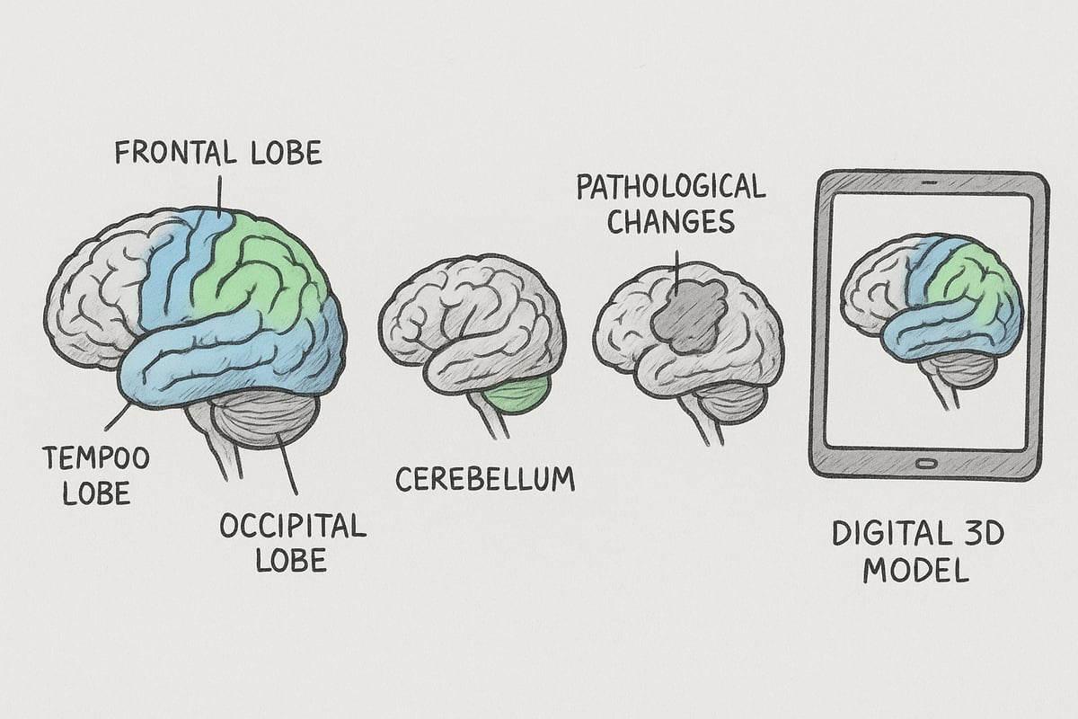
A classic anatomical brain model is often made in 1:1 size and divided into several segments. These models are popular in education as they provide a realistic picture of the structure of the brain. Many classic models have clear color coding that helps the user identify the different areas of the brain, such as the lobes, cerebellum and brainstem.
The segmentation makes it easy to separate and assemble the individual parts. It provides a tactile experience that strengthens the understanding of the brain's structure. The models are used primarily by students, teachers and health professionals in both practical and theoretical teaching.
Classic models often have labels that make it easy to learn the names and functions of the parts of the brain, making them an indispensable tool in any anatomy lesson.
Specialized models
Specialized models focus on selected areas of the brain, such as the cerebellum or brainstem. An anatomical brain model of this type allows for the study of specific regions in detail and can be indispensable in research or specialized teaching.
Some specialized models can be disassembled to examine blood vessels or nerve pathways separately. This provides a deeper understanding of functional relationships. There are also models where specific diseases or injuries are marked to illustrate clinical issues.
These models are often used in higher education or in clinical practice, where a high level of detail is crucial for learning outcomes.
Pathological and pedagogical models
An anatomical brain model can also be designed with a focus on pathological conditions. These can be models that show tumors, hemorrhages, or other disease changes. These models are a powerful tool in patient communication, as they make it easier to explain complex diagnoses and treatment options.
Educational models are often equipped with additional color codes or illustrations to support learning. They can be used in special education or to promote understanding in children and young people.
Using pathological models in teaching can increase understanding of disease processes and create greater confidence in patients under supervision.
Digital and interactive models
Digital developments have made it possible to work with interactive and 3D-printed models. A digital anatomical brain model can be visualized on a screen or via AR/VR technology, which provides new opportunities for experimental learning.
Digital models can be rotated, enlarged and adapted to individual needs. Statistics show that several Danish educational institutions use digital solutions as a supplement to physical models. The advantage of digital solutions is flexibility and access to updated knowledge.
However, many students miss the tactile experience that physical models provide when working with digital teaching.
Materials and durability
The choice of material has a great impact on both function and lifespan. An anatomical brain model is typically made of plastic, silicone or resin. Plastic models are lightweight and durable, while silicone gives a more lifelike feel. Resin is often used for advanced models where the level of detail needs to be extra high.
| Material | Benefits | Disadvantages |
|---|---|---|
| Plastic | Light, robust | Less lifelike |
| Silicone | Soft, realistic | Can be worn |
| Resin | High level of detail | More expensive, heavier |
Durability depends on proper maintenance and storage. If you choose a high-quality model, it can last for many years without losing precision.
Prices and availability as well as choice of model according to needs
The price of an anatomical brain model varies depending on size, level of detail and choice of materials. Basic models start around 1,000 DKK, while advanced research models can cost over 5,000 DKK. Availability in Denmark is high, and there are several suppliers who offer both standard and custom-made models.
When choosing a model, you should start with your needs. Students often benefit from compact, segmented models with clear color coding. Professionals and researchers may need more specialized solutions. If you are a student, you can find further guidance in ourGuide to Anatomy for Students .
The most important thing is to choose a model that matches both your professional level and your application, so that you get the most out of your investment.
How to Use an Anatomical Brain Model: Step-by-Step Guide
Working with an anatomical brain model requires a methodical approach, whether you are a teacher, student, or healthcare professional. This step-by-step guide will help you get the most out of your anatomical brain model and ensure that learning is both effective and engaging. Follow the next steps to gain a deeper understanding of the structure and function of the brain.
1. Preparation and setup
Start by choosing the anatomical brain model that best suits your purpose. Consider whether the model will be used for teaching, research, or patient guidance. Prepare all relevant materials, such as labels, manuals, and possibly anatomical posters and educational materials to support your presentation.
Place the model on a stable surface with good lighting. Use color-coded bases or accessories to highlight specific areas of the brain. Make sure all parts are intact and clean before starting.
One tip is to have a checklist ready so you don't forget important elements during the review. This way you ensure that your anatomical brain model is ready for use.
2. Introduction to the main parts of the brain
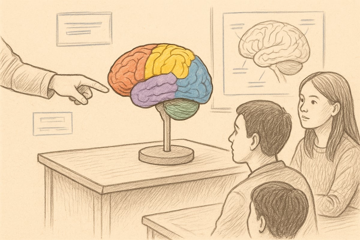
Begin by introducing the main areas of the model. Identify the cerebrum, cerebellum, brainstem, and the different lobes and blood vessels. Use labels and color coding to make it easier for participants to follow.
Demonstrate how the individual parts can be taken apart and put back together. This promotes understanding of the three-dimensional structure of the brain. Feel free to use a pointer or marker to point out areas along the way.
By introducing the main parts in this way, you help your participants form an overview of how an anatomical brain model reflects the real brain.
3. In-depth review of structures
Go deeper into the individual structures in the anatomical brain model. Start with the frontal lobe, explaining its role in cognition and movement. Continue with the parietal lobe, occipital lobe, and temporal lobe, highlighting their unique functions.
Use segmented models to show how the areas are connected. Also discuss the importance of blood vessels and the brainstem for vital functions.
Please provide examples with clinical cases where an injury in a specific area leads to specific symptoms. This makes the review both more practical and relevant.
4. Interactive learning and exercises
Actively involve participants by letting them work with an anatomical brain model along the way. Arrange small quizzes where they have to identify structures or describe functions. Group students and let them solve case-based tasks with the model as the focal point.
Experience shows that physically handling the model significantly increases learning outcomes. Feel free to combine the exercises with visual aids, such as posters or digital resources.
End with a short summary, asking what insights the students have gained by using the anatomical brain model hands-on.
5. Combined use with digital resources
Supplement the physical anatomical brain model with digital tools to create variety in your teaching. Integrate AR or VR so participants can explore the brain in a virtual environment. Use digital 3D models to show details that may be difficult to see on physical models.
Feel free to compare the two approaches and discuss their advantages and limitations. While the physical model provides tactile learning, digital resources provide dynamic perspectives.
A combination of both methods can provide a more complete understanding of the brain's complex anatomy.
6. Evaluation of learning outcomes
Evaluate the effectiveness of your teaching with an anatomical brain model by testing participants before and after use. Use short quizzes or practical tasks to measure understanding. Document the results to track progress over time.
Statistics show that using physical models improves both comprehension and memory compared to traditional reading alone. Consider collecting feedback from participants to continually improve your teaching.
Conclude by summarizing the most important points and highlighting how the model has strengthened the learning process.
7. Mistakes and pitfalls
Be aware of common mistakes when working with anatomical brain models. A common misconception is confusing the functions or locations of the lobes. Avoid separating model parts in a way that does not match the real anatomy.
Always follow the manufacturer's instructions, especially for complex models with many segments. Use both hands when moving larger models to avoid damage.
By focusing on correct use and maintenance, you minimize the risk of errors and ensure that your anatomical brain model remains a reliable tool for many years.
Areas of application: Education, research and clinic
Anatomical brain models are widely used in many contexts where understanding the structure of the brain is essential. Whether it is teaching, clinical work, research or dissemination, this model has a central role. Below we see how the model contributes to knowledge and learning in different environments.
In educational institutions
In schools, colleges and universities, the anatomical brain model is an indispensable tool for both teachers and students. The model makes it possible to visualize complex anatomical structures, which enhances understanding and facilitates learning. Medical students often use segmented models for exam preparation and group work.
According to an industry analysis, up to 80 percent of Danish medical students use physical models in anatomy teaching. The use of anatomical models in teaching helps to concretize theoretical knowledge and make it more accessible.
In the healthcare sector
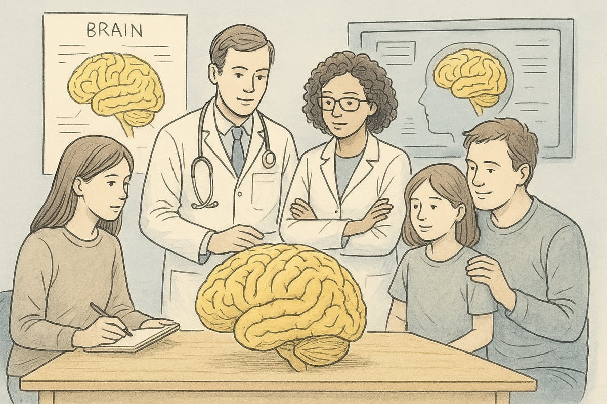
In the healthcare sector, anatomical brain models play an important role in patient communication and professional dissemination. Neurologists and other healthcare professionals use models to explain diseases, surgeries, or treatments to patients and relatives.
A model makes complex explanations more tangible, which increases the patient's understanding and confidence. Especially in neurological disorders, visual aids such as brain models can make a big difference to the dialogue between patient and therapist.
In research and innovation
In neuroscience research and the development of new treatment methods, anatomical brain models are of great importance. Scientists use detailed models to simulate surgical procedures and analyze the structure of the brain.
The model makes it possible to test hypotheses and try out techniques before they are implemented in practice. In medical innovation, advanced models are also used to develop new procedures and tools, making them central to both research and development.
In public information and museum communication
Anatomical brain models are used not only in professional environments, but also in public education. In museums and exhibitions, models are included as interactive elements where visitors can explore the structure of the brain.
These models help make scientific knowledge accessible to a wider audience. Educational events and lectures often use brain models to engage and inspire both children and adults to learn more about the brain.
Home use and hobby
Individuals with an interest in anatomy or art often choose to invest in an anatomical brain model for home use. The model can serve as a decorative object, study object, or inspiration for creative projects.
For hobbyists, artists and students, the model offers the opportunity to explore the details of the brain and delve into its structure on their own terms. This makes the brain model accessible to anyone with a curiosity about anatomy.
Benefits for different user groups
The benefits of an anatomical brain model vary depending on the user group:
| User group | Advantage |
|---|---|
| Teachers | Better communication and engagement |
| Student | Increased understanding and better exam results |
| Healthcare professionals | Effective patient communication |
| Researchers | Precise simulation and analysis |
| Private individuals | Personal learning and inspiration |
Regardless of the area of application, the model creates value by making the complex structure of the brain more understandable and present.
How to choose the right anatomical brain model
Choosing the right anatomical brain model requires careful consideration of needs, quality, and application. To ensure optimal learning and understanding, it is important to review a number of criteria before making your decision.
Assess your needs and objectives
Start by defining the primary purpose of your anatomical brain model. Will it be used for teaching, research, patient guidance, or perhaps as a hobby? A model for detailed teaching at the university has different requirements than one used for quick patient information in the clinic.
Think about the depth of knowledge you want to convey. If you work with neurophysiology or functional anatomy, a course likeFunctional Anatomy and Neurophysiology can provide inspiration for choosing the type of model and level of detail.
Important selection criteria
When choosing an anatomical brain model, you should focus on several key aspects:
- Size: Choose between 1:1, 2x or miniature depending on space and needs.
- Segmentation: Consider whether the model should be separable into parts for better learning.
- Color coding and labels: Easy recognition of structures increases learning outcomes.
- Material: Plastic, silicone or resin each have their own advantages and disadvantages.
A quick overview can be summarized in the table below:
| Criterion | Advantage | Disadvantage |
|---|---|---|
| Segmentation | Possibility of separation | Can be fragile |
| Color coding | Increases clarity | May seem artificial |
| Silicone | Soft and realistic | Less durable than plastic |
Price and budget
The price level for an anatomical brain model varies considerably. Basic models start around 1,000 DKK, while advanced research models can cost over 5,000 DKK. Consider your budget in relation to purpose and frequency of use.
It is often worth investing in a higher quality model if it is to be used intensively or for detailed teaching, but for students a cheaper alternative may be sufficient.
Quality and durability
The quality of an anatomical brain model depends primarily on the choice of materials, assembly, and level of detail. Models from reputable manufacturers often offer greater precision and longer durability.
Consider whether the model comes with a warranty and whether repairs or spare parts are available. A robust model ensures that your investment and learning outcomes last for many years.
Availability and delivery
It is crucial to choose a supplier with proven expertise in anatomical brain models and good customer service. Specialized retailers like Om eAnatomi and our expertise offer both advice, fast delivery and access to a wide selection.
Always check delivery times and the availability of support and guidance during the purchasing process. This contributes to a safe and efficient purchase.
Possibility of customization
Some suppliers offer custom solutions where your anatomical brain model can be customized with additional accessories or custom-made parts. This is particularly relevant for research projects or specialized teaching courses.
Choosing a model with customization options can give you flexibility if your needs change over time.
Recommendations from users and experts
Before making your final choice, it is recommended to read reviews from users, educators, and clinicians. Experience with clear color coding, segmentation, and durability can help you choose the anatomical brain model that best meets your requirements.
A model that is praised for its ease of use and accuracy will often provide greater satisfaction and learning outcomes in the long run.
Maintenance and storage of anatomical brain models
Ensuring proper maintenance and storage of an anatomical brain model is essential for both functionality and longevity. With proper care, the model will maintain its accuracy and neat appearance for many years. Follow the advice below to get the most out of your anatomical brain model.
Cleaning and hygiene
An anatomical brain model should be cleaned regularly with mild detergents, especially if used in patient guidance. Avoid using harsh chemicals as they can damage the surface. Use a soft cloth and lukewarm water for plastic and silicone. Dry the model thoroughly after cleaning to avoid moisture damage.
Storage
For optimal durability, an anatomical brain model should be stored dry and away from direct sunlight. It is recommended to use the original box or a dust-proof container. Statistics show that proper storage can extend the life of the model by up to 5 years. Avoid humid environments and extreme temperatures.
Repair and spare parts availability
If an anatomical brain model consists of segmented parts, individual components may become loose or damaged. Most suppliers offer spare parts or repairs so that the model can be quickly returned to use. Replacing small parts is often simple and ensures continued functionality.
Safe handling
Always handle your model with care. Lift larger models with both hands and avoid dropping them. Impacts and knocks can cause permanent damage or loss of detail. Place the model on a stable surface when not in use.
Documentation and manuals
Always keep the instructions and documentation that came with the model. Manuals often contain detailed illustrations that facilitate proper use and assembly. This ensures that parts are assembled correctly and the model is used correctly over time.
Environmental considerations
When an anatomical brain model reaches its end of life, it should be disposed of in an environmentally friendly manner. Many models can be disassembled so that certain materials can be recycled. Ask the supplier about options for recycling or proper waste management to minimize environmental impact.
Now that you have an overview of how anatomical brain models can bring learning, teaching and patient communication to life, you can probably see the value in choosing the right model for your needs. Whether you are a student, teacher or healthcare professional, an accurate and well-designed brain model can give you a significantly better starting point for understanding the complexity of the brain. If you would like to explore the wide range of models developed in collaboration with experts and adapted for different purposes, you can Read more here .
0 comments