

Discover how a 3D anatomical model can change your approach to anatomy and health science in 2025.
This guide gives you insight into how to choose and use a 3D anatomical model optimally, whether you are teaching, researching or working clinically.
We explore the latest technologies, selection criteria, pedagogical benefits, practical examples, and the most important trends for the future.
Get inspired to take the next step and discover how a 3D anatomical model can take your understanding and teaching to a new level.
What is a 3D anatomical model?
A 3D anatomical model is today an indispensable tool in health education, research and clinical practice. The model makes it possible to visualize and understand the structures of the body in a way that cannot be achieved with traditional illustrations. The development has led to a wide range of models, both physical and digital, covering all aspects of anatomy.
Definition and basic principles
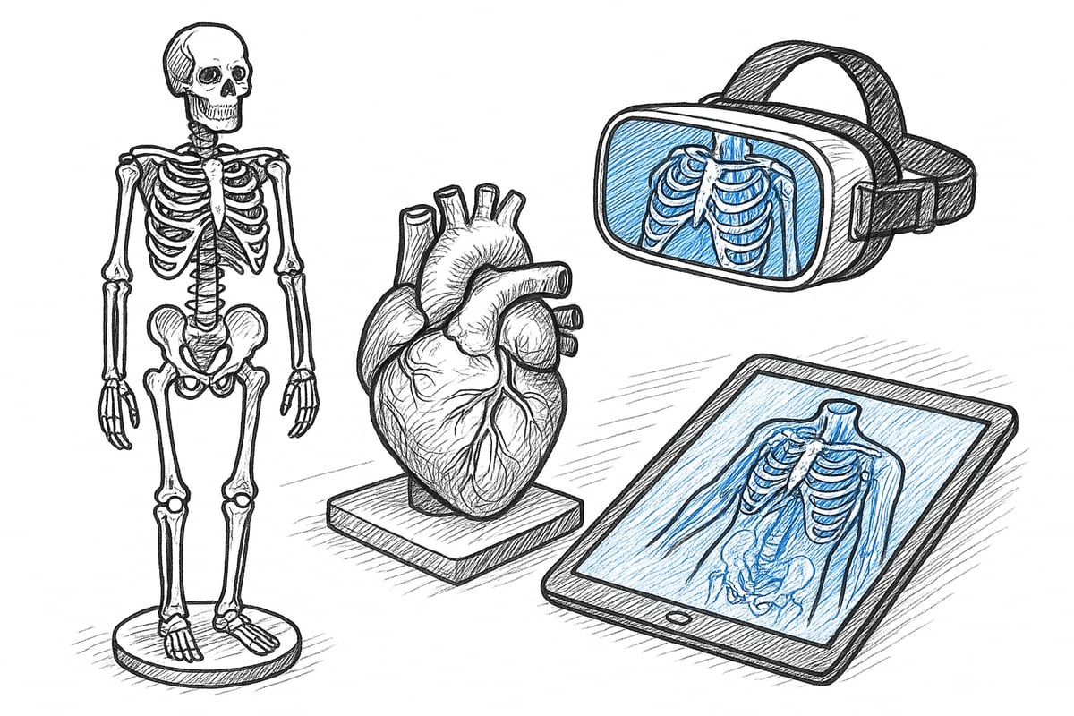
A 3D anatomical model is a three-dimensional representation of human or animal anatomy, designed to illustrate the body's structures in detail. The models are available both as physical objects and digital solutions.
Physical models are often made of plastic, resin or silicone. They can be realistic and durable, and allow for tangible interaction. Digital models, on the other hand, are experienced through computers, tablets or VR/AR technologies, opening up virtual experiences and simulations.
The purpose of a 3D anatomical model is to provide a spatial and detailed image of anatomy. The models are widely used for teaching, training and research. Throughout history, development has moved from simple wax models to advanced digital solutions, which today set new standards for learning.
If you are a student and want practical guidance in the use of models, you can find inspiration atFor students about anatomical models .
Types of 3D anatomical models
There are many types of 3D anatomical models, adapted to different purposes and fields of study. The most common are:
- Human models: Skeleton, organs, muscles, nerves, etc.
- Veterinary models: Anatomy of animals such as dogs, horses and cats.
- Microscopic models: Cells and tissues in enlarged format.
- Interactive digital models: VR/AR applications where the user can explore the body virtually.
Universities and hospitals often use specialized models, for example for surgical training or case-based learning. The digital revolution means that more institutions are choosing virtual models, especially for distance learning.
Technological advances in 2025
In 2025, the market for 3D anatomical models will be characterized by rapid technological advances. 3D printing technology has made it possible to produce models with great precision and adapted to individual needs. This applies to both teaching models and patient-specific solutions.
The integration of augmented reality (AR) and virtual reality (VR) has become standard in many educational institutions. Statistics show an increase of over 30 percent in the use of digital models from 2022 to 2025. Popular software solutions such as Visible Body and 3D Organon allow the user to explore anatomy interactively and realistically.
The development creates new opportunities for learning and training, where technology strengthens understanding and makes teaching more engaging.
Advantages of 3D over 2D
A 3D anatomical model offers significant advantages over traditional 2D illustrations. The most important benefit is better spatial understanding, as the model can be viewed and rotated from all angles.
Interactive learning is a great advantage, especially when the model can be disassembled or explored digitally. Patient communication is improved because complex topics can be clearly visualized.
International studies document that the use of a 3D anatomical model can increase learning outcomes by up to 50 percent, making the model an effective tool for students, educators, and healthcare professionals alike.
Disadvantages and limitations
Although 3D anatomical models offer many advantages, there are also challenges. The price of advanced models can be high, especially for digital solutions with VR/AR.
Technical equipment is often required, which may limit accessibility in certain classrooms or institutions. Digital models must be continuously updated and maintained to ensure relevance and functionality.
For example, implementing new technologies can be met with resistance from users who are not familiar with digital platforms. It is therefore important to consider needs, budget and technical resources before investing in a 3D anatomical model.
How to choose the right 3D anatomical model
Choosing the optimal 3D anatomical model requires careful consideration. The purpose, user needs and practical framework should always be the starting point. A systematic approach simplifies the process and ensures that the investment will support learning, research or clinical application.
Purpose and target group
Start by defining who will use your 3D anatomical model. Will it be used at the elementary school level, university or continuing education? Different fields such as medicine, physiotherapy, veterinary or biology have varying requirements for level of detail and functionality.
For example, medical students need models with high anatomical accuracy, while biology classes in elementary schools can make do with more general structures. Also consider whether the model will be used for theoretical teaching, practical training, or patient communication.
Materials and quality
The choice of material has a big impact on how realistic and durable your 3D anatomical model is. The most common materials are plastic, resin, silicone and 3D printed polymers. Each type has advantages and disadvantages:
| Material | Realism | Durability | Price level |
|---|---|---|---|
| Plastic | Low | High | Low |
| Resin | High | Between | Between |
| Silicone | Very high | Between | High |
| 3D printing | Variable | Variable | Falling |
Make sure the model has relevant certifications such as CE or ISO, especially if it is to be used in the healthcare sector.
Functionality and interactivity
Consider what features your 3D anatomical model should have. Some models can be disassembled and reassembled, allowing for active learning and immersion in complex structures.
Digital models often offer VR or AR integration as well as app control. Popular interactive models can visualize blood vessels, nerves or microscopic details in real time, enhancing the teaching experience.
Consider whether physical or digital interactivity best matches your learning goals and usage scenarios.
Price and budget considerations
The price of a 3D anatomical model varies greatly depending on material, size and functionality. Physical models typically have a higher initial cost, while digital solutions require investment in software and hardware.
It is important to consider the total cost over time, including maintenance and updates. Statistics show that the price of 3D printed models has decreased by 20 percent in the past three years, making advanced models more accessible.
Always compare several offers and consider which solution provides the most value for money.
Availability and suppliers
When purchasing a 3D anatomical model, you should research the market thoroughly. Reputable suppliers can advise on the right choice for your specific needs.
Ask questions about warranty, update options, and technical support. Institutional purchases can often get discounts and access to customized solutions, which is a benefit for both schools and hospitals.
Also check the suppliers' experience with the education sector and their ability to provide documentation and certifications.
eAnatomi ApS: Danish expert in anatomical models
eAnatomi ApS is a leading Danish specialist in 3D anatomical models and educational materials. The company works closely with healthcare experts to ensure that all models and posters are scientifically accurate.
The selection is wide-ranging: From models for students and educators to clinical solutions for hospitals and private individuals. Original designs and the option of customized models are offered, so that all customer segments have their needs met. Read more about the company's background and expertise at About eAnatomi and expertise in models .
Educational and professional benefits of 3D anatomical models
3D anatomical models have changed the way we understand and communicate anatomy. Modern technologies make it possible to visualize complex structures in much greater detail, enhancing both learning and clinical practice. Here we review the most important pedagogical and professional benefits of using 3D anatomical models today.
Improved learning and understanding
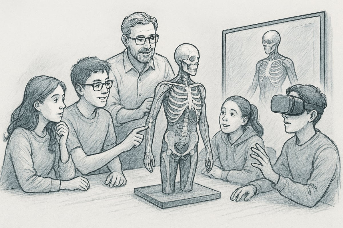
Studies show that the use of 3D anatomical models significantly increases learning outcomes compared to traditional 2D illustrations. Medical and physiotherapy students gain a deeper understanding of the body's complex structures when they can explore them physically or digitally.
Visualizing organs, muscles, and the skeleton in three dimensions makes it easier to understand connections and relationships. This is especially true in areas where detail and spatiality are crucial to mastering the subject.
Many educators find that the transition to 3D anatomical models strengthens students' self-confidence and ability to apply theoretical knowledge in practice. This makes the models an indispensable tool in modern healthcare education.
Active learning and engagement
Hands-on interaction with a 3D anatomical model promotes active participation and engagement in teaching. When students are given the opportunity to assemble and disassemble models, both motivation and learning efficiency increase.
Simulation-based teaching, where models are used to illustrate scenarios or train specific skills, has proven particularly effective. Statistics show that 70 percent of students prefer 3D anatomical models to flat images.
For educators who want to integrate models into their practice, there are resources and guides, such as For Educators: Effective Use of Models , which provides concrete examples and tips for optimal use.
Clinical use and patient communication
In clinical practice, 3D anatomical models play a central role for both healthcare professionals and patients. The models make it easier to explain disease processes, treatments and anatomical conditions, so that the patient better understands their situation.
In surgical planning and preoperative training, a 3D anatomical model can be used to simulate procedures and visualize patient-specific challenges. Especially in orthopedic surgery, models are used to tailor interventions and minimize risks.
Patients experience greater security when they are presented with diagnoses and treatments visually. This strengthens communication and increases the chance of a positive treatment outcome.
Skills training and simulation
A 3D anatomical model makes it possible to train procedures and techniques without risk to the patient. This applies to, for example, injection training, surgical simulation, or exercises in identifying anatomical structures.
Data shows that the use of 3D anatomical models reduces errors in training environments by up to 40 percent. This is because repeated practice on realistic models provides better muscle memory and increased safety.
Training sessions with models can be adapted for both beginners and experienced users, making them relevant across educational levels and specialties.
Interdisciplinary application
The benefits of 3D anatomical models extend beyond medicine and physiotherapy. The models are used in veterinary education, dentistry education and biology, where they support the understanding of both human and animal anatomy.
Collaboration between different disciplines is strengthened when everyone works from the same visual foundation. Statistics from 2022 to 2025 show a 25 percent increase in the use of 3D anatomical models in the veterinary sector.
Universities and institutions report that interdisciplinary development projects with models increase innovation and lead to better learning tools for all parties.
Accessibility and inclusion
3D anatomical models support students with different learning styles, as visual, auditory and kinesthetic elements can be incorporated into the teaching. Digital models also enable distance learning and hybrid solutions.
During the COVID-19 pandemic, many institutions experienced great success with online platforms where students could interact with 3D anatomical models from home. This has made teaching more flexible and inclusive.
The models therefore contribute to increased accessibility and equal opportunities for everyone, regardless of geographical location or special needs.
Step-by-step guide: How to implement 3D anatomical models
Implementing a 3D anatomical model in teaching or clinical practice requires a structured approach. By following this guide, you can ensure that both educators and students get the most out of the technology. Each step below will help you create the best framework for effective use and learning.
Step 1: Needs analysis and goal setting
Before choosing a 3D anatomical model, analyze your needs. Start by identifying learning objectives and the specific use case. Is the goal to improve the understanding of human anatomy for medical students, or will the model be used for training in clinical procedures?
Involve relevant educators, managers and users in the process. This ensures that the model matches both the curriculum and practical requirements. For example, a medical faculty can prepare a list of curriculum needs and desired features so that the investment in a 3D anatomical model is targeted and effective.
Step 2: Selection and procurement
Once your needs are clear, compare different models based on quality, functionality and price. Get quotes from multiple suppliers and research which models best support your goals.
Focus on warranty, support and the possibility of updates. Remember that the overall benefit of a 3D anatomical model also depends on how easily it can be integrated into existing teaching environments. Use a table to compare models on parameters such as durability, interactivity and price.
| Parameters | Model A | Model B | Model C |
|---|---|---|---|
| Durability | High | Between | High |
| Interactivity | Digital + physical | Physical | Digital |
| Price | Medium | Low | High |
Step 3: Integration into teaching or clinical practice
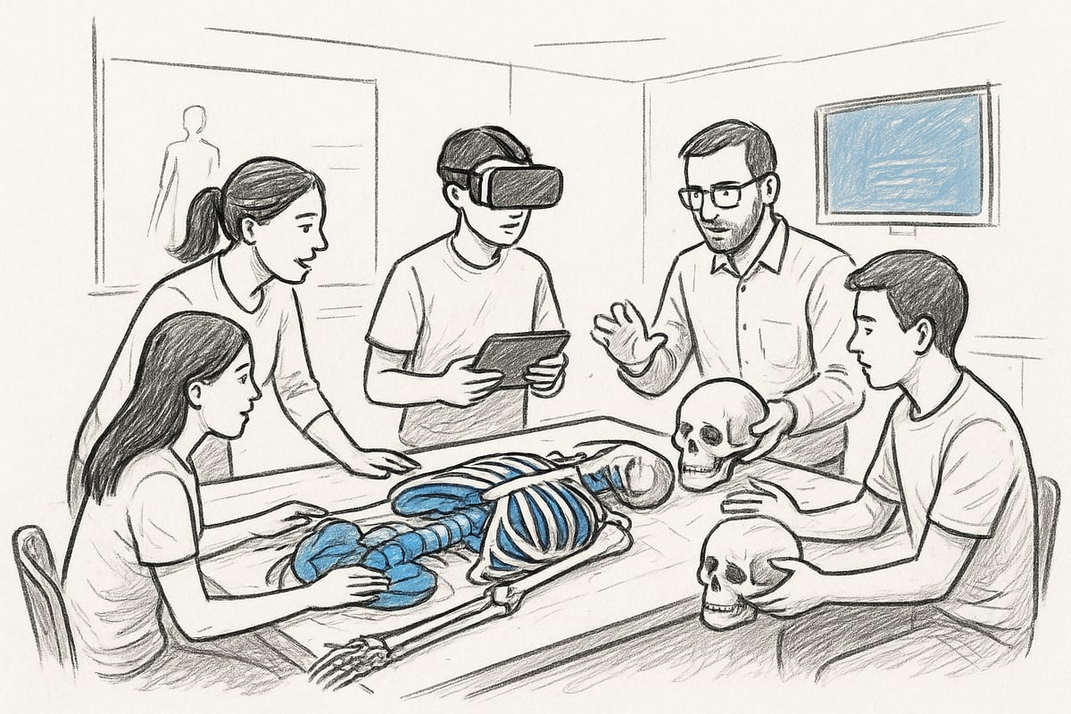
Develop lesson plans or training programs that actively incorporate 3D anatomical models. Combine the model with existing materials to enhance learning. Consider hybrid teaching, where both physical and digital models are used side by side.
Digital solutions open up new possibilities, and with VR or AR you can create engaging learning environments. A good complement is to explore resources like Transforming Education: Integrating AI, AR, VR, and 3D Printing for Effective Learning , which provides practical knowledge on integrating advanced technologies into teaching.
Step 4: Training users
To ensure optimal utilization of a 3D anatomical model, users must receive thorough training. Hold workshops and introductory courses where both teachers and students have the opportunity to try the model in practice.
Develop user manuals and guides so that everyone feels comfortable with the technology. Statistics show that 80% faster adaptation is achieved when the training is structured and tailored to the target group. This is especially true if the implementation includes digital solutions or VR technology.
Step 5: Ongoing evaluation and updating
Systematically collect feedback from both users and students. Evaluate how the 3D anatomical model works in practice and identify any needs for updating models or software.
Many institutions conduct annual evaluations where experiences are shared and future needs are mapped. This ensures that you are always working with the most relevant and up-to-date models and that your investment continues to provide value.
Step 6: Maintenance and support
A 3D anatomical model requires ongoing maintenance to maintain functionality. Establish routines for cleaning and proper storage of physical models. For digital solutions, there should be access to technical support and updates.
Consider service agreements with vendors so that any issues can be resolved quickly. A well-run support structure minimizes downtime and ensures that both teaching and clinical training can continue without interruption.
Future trends for 3D anatomical models
The future of 3D anatomical modeling is rapidly advancing thanks to groundbreaking technologies and new forms of collaboration. These developments are creating opportunities that make it easier to adapt education, research, and clinical practice. Here is an overview of the six most important trends shaping the use of 3D anatomical modeling in 2025.
Development within 3D printing and materials
3D anatomical models have seen significant advances in material selection and printing technology in recent years. New biocompatible and sustainable materials make it possible to produce models that are both more realistic and environmentally friendly.
With faster and more accurate 3D printers, detailed structures that were previously impossible to produce can now be created. According to industry analysts, the 3D printing market in medical education is expected to grow by 15 percent annually, underscoring the increasing demand for advanced 3D anatomical models.
Integration of AI and machine learning
Artificial intelligence has begun to revolutionize the development of 3D anatomical models. With AI, it is now possible to automate the adaptation of models to individual patients, making them more relevant for precision medicine and advanced training.
Furthermore, machine learning enables faster diagnosis and more effective skill training. For further insight, read the research paper Generative AI Framework for 3D Object Generation in Augmented Reality , which demonstrates how AI can generate 3D objects in real time and integrate them into AR environments. This development makes 3D anatomical models even more interactive and user-friendly.
Expanded use of VR and AR
Virtual and augmented realities have changed the way we work with 3D anatomical models. Several institutions are now using VR to create virtual classrooms and clinical simulations, increasing the accessibility and quality of teaching.
A recent study shows that 35 percent more institutions will use VR in anatomy education by 2025 than before. Read more about innovative solutions in the study Holoview: Interactive 3D Visualization of Medical Data in AR , which illustrates how AR can transform interactive learning. This makes 3D anatomical models an even more powerful tool for both students and professionals.
Global accessibility and digitalization
Digitalization has made it easier than ever to access 3D anatomical models globally. With cloud-based solutions and online platforms, educators and students can work with models regardless of geographic location.
Statistics show that 60 percent of universities now offer digital 3D anatomical models, strengthening the possibilities for distance learning and collaboration across borders. This development supports both inclusion and flexibility in learning environments.
Individualized and patient-specific models
One of the most exciting trends is the creation of 3D anatomical models based on patients' own scans. This opens up applications in precision medicine, where orthopedic surgeons and oncologists can plan treatments with models that match the individual patient exactly.
This individualization improves both diagnosis and treatment. 3D anatomical models are thus becoming an important tool in modern healthcare, where tailored solutions are in focus.
Collaboration between research, education and industry
The development of 3D anatomical models is further accelerated through partnerships between universities, companies and hospitals. Cross-sectoral projects ensure faster innovation and access to the latest technologies.
Examples of such collaborations are joint development projects where researchers and educators work together to create better learning tools. The future of 3D anatomical models lies in this interaction, where knowledge and technology are united for the benefit of all users.
Once you have an overview of the many possibilities and benefits of 3D anatomical models, it can be difficult to know where to start. Whether you work in teaching, research or clinical practice, the right model can make a significant difference to both learning and communication. At eAnatomi ApS you will find a wide selection of models and posters that have been developed together with healthcare experts and tailored to your needs. If you want to delve deeper into the selection or need some sparring for your next project, you can Read more here
0 comments