
Varför är det så viktigt att förstå människans anatomi i dagens utbildnings- och hälsovårdsmiljö? En anatomisk modell med organ ger studenter, lärare och vårdpersonal ett ovärderligt verktyg för att visualisera och lära sig om kroppens struktur. Den här guiden ger dig en översikt över de olika typerna av modeller, deras användningsområden, hur du väljer rätt lösning och hur du underhåller modellerna. Upptäck hur dessa modeller kan göra en verklig skillnad i lärande, utbildning och kommunikation år 2025. Läs vidare och lär dig mer om dina alternativ.
Vad är en anatomisk modell med organ?
En anatomisk modell med organ är ett oumbärligt verktyg för alla som vill förstå människokroppens struktur i detalj. Denna typ av modell skiljer sig från allmänna anatomiska modeller genom att fokusera specifikt på de inre organen och deras relationer.
Modellerna tillverkas vanligtvis av material som plast, silikon, harts eller genom 3D-utskriftsteknik. Plastmodeller är hållbara och lätta att rengöra, medan silikon ger en mer realistisk känsla. Harts används ofta för detaljerade modeller, medan 3D-utskrift möjliggör produktion av individuella eller patientspecifika lösningar.
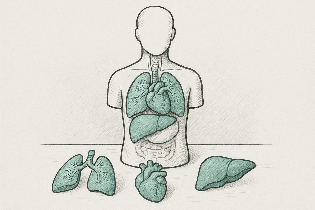
De varierar i storlek från små bordsmodeller till fullskaliga människokroppar. De mest populära är torsomodeller med avtagbara organ, vilket gör det enkelt att undersöka varje organs placering och funktion. Andra modeller fokuserar på bara ett organ, såsom hjärtat eller levern.
Syftet med en anatomisk modell med organ är att visualisera komplexa anatomiska strukturer och skapa möjligheter till praktiskt lärande. Modellen används främst för undervisning, träning och patientkommunikation. Många studenter upplever att lärandet förbättras avsevärt när de har möjlighet att fysiskt arbeta med en modell. Om du som student vill lära dig mer om hur modellerna används i praktiken kan du hitta inspiration på För studenter i anatomi .
Definition och grundläggande egenskaper
En anatomisk modell av organ är en fysisk representation av människokroppens inre organ, ofta i naturlig storlek. Dessa modeller är utformade för att illustrera organens placering, struktur och relationer.
Typiska material inkluderar:
- Plast: robust och hållbar.
- Silikon: mjuk och realistisk textur.
- Harts: exakt detaljnivå.
- 3D-utskrift: möjlighet till individualisering.
Storlekarna varierar från kompakta bordsmodeller till fullskaliga modeller, beroende på behov. Vanliga exempel är:
- Torso med avtagbara organ
- Hjärt-, lever- eller lungmodeller
- Modeller av matsmältningssystemet
Syftet med en anatomisk modell med organ är att låta användaren se, röra vid och förstå organens struktur. Detta underlättar inlärningen och gör komplexa ämnen mer konkreta. Modellen används flitigt inom både utbildning, patientinformation och yrkesutbildning.
Historisk utveckling och användning
Användningen av anatomiska modeller av organ har en lång historia som går tillbaka till vaxmodellerna från 1700-talet. Vid den tiden skapades modellerna för att hjälpa läkare och studenter att förstå kroppens insida utan att behöva utföra dissektion.
I takt med att tekniken utvecklades blev plast och senare silikon och harts populära material, vilket gjorde modellerna mer hållbara och detaljerade. Idag används 3D-utskrift, vilket möjliggör ännu högre precision och anpassning till specifika utbildningsbehov.
Fysiska modeller har blivit allt viktigare inom medicinsk utbildning. En rapport från 2023 visade att 75 % av läkarutbildningarna i Europa använder fysiska modeller som en regelbunden del av sin undervisning. Detta har förbättrat kvaliteten på anatomiundervisningen, eftersom modellerna gör det lättare att förstå rumsliga relationer och organfunktioner.
Ur ett pedagogiskt perspektiv bidrar en anatomisk modell med organ till ökat engagemang och läranderesultat. Både elever och lärare upplever att konkret visualisering underlättar förståelsen, särskilt i tvärvetenskapliga sammanhang.
Fördelar och nackdelar med anatomiska modeller med organ
En anatomisk modell med organ ger en noggrann och realistisk visualisering av kroppens inre strukturer. Detta gör det möjligt att öva på procedurer och få förståelse utan risk för patienterna. Modellen kan användas om och om igen, vilket är en stor fördel i undervisning och träning.
Fördelar inkluderar:
- Tydlig visualisering av komplexa strukturer
- Säker och repeterbar träning
- Praktisk inlärning som stärker förståelsen
Det finns dock också nackdelar. Priset för en anatomisk modell med organ kan vara högt, särskilt för avancerade eller specialdesignade modeller. De kräver utrymme för förvaring och måste underhållas ordentligt för att hålla sig snygga och funktionella.
Jämfört med digitala lösningar som appar eller VR erbjuder fysiska modeller en taktil upplevelse men saknar de interaktiva möjligheter och den flexibilitet som digitala verktyg kan ge. I praktiken kombinerar många institutioner båda typerna för att uppnå bästa möjliga inlärningseffekt.
De möjliga tillämpningarna sträcker sig brett, från klassrum och kliniker till museer och utställningar, där de bidrar till spridning av hälsokunskap på alla nivåer.
Typer av anatomiska modeller med organ
Utbudet av anatomiska organmodeller som finns tillgängliga idag är stort och mångsidigt. Olika typer av modeller täcker olika behov, från klassisk undervisning till avancerad medicinsk utbildning. Varje typ har sina egna styrkor och tillämpningsområden. Det är viktigt att känna till skillnaderna om du vill välja rätt anatomisk organmodell för din undervisning, klinik eller forskning.
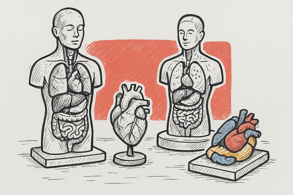
Klassiska modeller: Torso- och helkroppsmodeller
Den klassiska anatomiska modellen med organ inkluderar både torsos och helkroppsmodeller. En torsomodell visar ofta överkroppen med organ som kan tas bort och undersökas individuellt. Detta gör det möjligt för eleverna att utforska de inre organens placering och struktur genom fysisk interaktion.
Helkroppsmodeller täcker hela kroppen och används särskilt i avancerad undervisning där detaljerad kunskap om både ytliga och djupare strukturer krävs. Dessa modeller visar ofta muskler, skelett och organ i samspel. Populära modeller har färgkodade delar så att användarna enkelt kan identifiera organen. Att använda anatomiska modeller med organ i denna kategori ger en solid grund för att förstå människokroppen.
Organspecifika modeller
Organspecifika modeller fokuserar på ett enda organsystem, såsom hjärta, lungor, lever eller njurar. De är idealiska för undervisning där en djupgående förståelse av ett specifikt organ krävs. Till exempel låter en detaljerad hjärtmodell dig gå igenom blodets väg genom kamrarna och klaffar.
Lungmodeller kan illustrera lungvävnad, bronker och alveoler, medan levermodeller belyser gallgångarna och blodförsörjningen. Organspecifika anatomiska modeller med organ används ofta inom specialiserade discipliner som kardiologi, gastroenterologi eller nefrologi. De stöder både teoretisk kunskap och praktisk insikt.
Funktionella och interaktiva modeller
Funktionella anatomiska modeller med organ har rörliga delar eller elektroniska komponenter som gör inlärningen mer aktiv. Till exempel kan vissa matsmältningsmodeller visa peristaltiska rörelser för att göra processen lättare att förstå. Andra modeller har inbyggda lampor eller ljud som demonstrerar blodflöde eller hjärtslag.
Dessa interaktiva modeller möjliggör simulering av kliniska situationer och övning av procedurer utan risk. De används ofta i simuleringsträning för läkarstudenter eller vårdpersonal. Fördelen är att användaren får praktisk erfarenhet och kan upprepa övningar tills förståelsen är etablerad.
3D-utskrivna modeller och kundanpassade lösningar
Den senaste utvecklingen inom anatomiska organmodeller är 3D-utskrivna och patientspecifika modeller. 3D-utskrift gör det möjligt att skapa modeller från skanningar av riktiga patienter, vilket ökar precisionen och möjligheten till individualisering.
Dessa modeller används främst för kirurgisk planering och avancerad utbildning, där subtila variationer i anatomin kan vara betydande. Användningen av 3D-utskrivna medicintekniska produkter förväntas öka med 35 procent från 2022 till 2024. Anpassade lösningar kan också anpassas för undervisningsändamål där specifika sjukdomsmönster eller skador ska illustreras. Anatomiska modeller med organ i denna kategori ger unika möjligheter till personligt och effektivt lärande.
Anatomiska affischer och digitala bilagor
Anatomiska modeller med organ kompletteras ofta av affischer och digitala verktyg. Affischer ger en visuell översikt över kroppens system, medan digitala lösningar som appar och VR erbjuder interaktivt lärande.
Kombinationen av fysiska modeller och digitala hjälpmedel förbättrar undervisningen och gör det lättare att förstå komplexa ämnen. Många lärare väljer att använda båda för att tillgodose olika inlärningsstilar. För inspiration till danska affischer, se t.ex. Anatomiska affischer designade i Danmark , som erbjuder pedagogiska och estetiska lösningar för klassrummet eller kliniken. Detta gör anatomiska modeller med organ ännu mer användbara och flexibla i framtidens undervisning.
Användning av anatomiska modeller med organ
Användningen av en anatomisk modell med organ varierar mycket, från klassrum till klinik och museum. I det här avsnittet fördjupas i hur modellerna används i olika miljöer för att förbättra lärande, utbildning, patientförståelse och kommunikation.
Utbildningssektorn: Skolor, högskolor och universitet
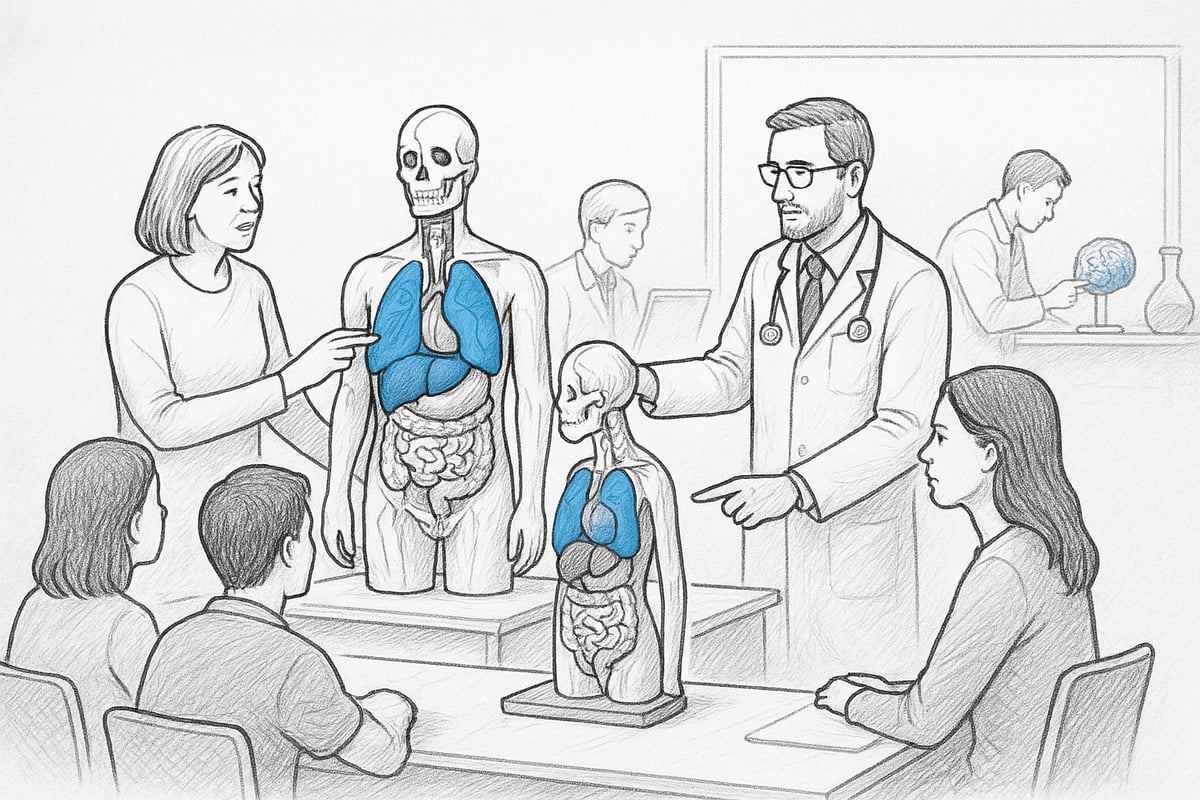
Inom utbildningssektorn är en anatomisk modell med organ ett oumbärligt verktyg. Biologi och hälsoundervisning blir mycket mer konkret när elever och studenter fysiskt kan undersöka och sätta ihop organen. Detta stärker förståelsen för kroppens struktur och funktion.
Studier visar att användningen av fysiska modeller kan förbättra provresultaten avsevärt. En studie dokumenterade upp till 30 % bättre resultat hos elever som aktivt arbetar med modeller jämfört med rent teoretisk undervisning. Den praktiska dimensionen stöder både visuellt och taktilt lärande.
Om du vill fördjupa dig i hur modeller används i undervisning kan du läsa mer i den här artikeln: Anatomiska modeller i undervisningen .
Sjukvårdspersonal och klinisk utbildning
För vårdpersonal är en anatomisk modell med organ avgörande i utbildningen av läkare, sjuksköterskor och terapeuter. Modellerna gör det möjligt att öva på procedurer och diagnostiska tekniker i en säker miljö utan risk för patienter.
Simuleringsträning med modeller skapar en realistisk inlärningsmiljö där upprepning och misstag är tillåtna. Detta ökar både vårdpersonalens självförtroende och kompetens, och bidrar till bättre behandlingskvalitet i praktiken.
Praktiska övningar med fokus på organsystem, såsom hjärta eller lungor, kan differentieras efter specialisering och nivå.
Patientutbildning och kommunikation
En anatomisk modell med organ är också ett kraftfullt verktyg i dialogen mellan vårdpersonal och patienter. Modellerna används för att illustrera diagnoser, procedurer och behandlingsförlopp, vilket gör komplex information mer lättillgänglig.
När patienter ser och rör vid en modell ökar deras förståelse och engagemang, vilket ofta leder till bättre följsamhet och mer konstruktiva samtal om sjukdomen och behandlingen.
Modeller kan också användas för att avmystifiera kroppen för barn och unga, vilket skapar trygghet och nyfikenhet.
Forskning och utveckling
Inom forskning och utveckling används anatomiska modeller med organ för att testa och utveckla medicintekniska produkter och nya kirurgiska tekniker. Modellerna skapar en säker miljö för experiment utan risk för levande vävnad.
Samarbeten mellan universitet och sjukhus gör det möjligt att utveckla patientspecifika modeller som kan användas för exakt planering av komplexa procedurer. Modellerna kan anpassas till individuella anatomiska variationer med hjälp av 3D-utskrift.
Denna metod främjar innovation och bidrar till mer precisa och effektiva behandlingar, vilket i slutändan gynnar patienterna.
Andra användningsområden: Utställningar, museer och offentlig verksamhet
En anatomisk modell med organ är också ett populärt kommunikationsverktyg i utställningar och museer. Här används modellerna för att levandegöra hälsovetenskaplig kunskap för både barn och vuxna.
Gästerna kan interagera med modellerna, vilket gör komplexa ämnen lättare att förstå och komma ihåg. Skolor besöker ofta museer med anatomiska utställningar för att stärka elevernas intresse för naturvetenskap.
Modellerna används också i folkhälsokampanjer, där de hjälper till att förklara vikten av organfunktion och hälsosamma livsstilar på ett visuellt och engagerande sätt.
Hur man väljer rätt anatomisk modell med organ
Att välja rätt anatomisk organmodell kräver insikt i både användarnas behov och de många alternativen på marknaden. Nedan går vi igenom hur du bäst gör ett välgrundat val för att få ut det mesta av din investering.
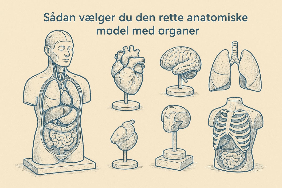
Syfte och målgrupp
Det första steget är att definiera syftet med din anatomiska organmodell. Kommer den att användas för grundläggande biologiundervisning, avancerad medicinsk utbildning eller patientinformation?
Målgruppen är mycket viktig. En anatomisk modell med organ för grundskoleelever måste vara robust och pedagogiskt enkel, medan modeller för universitet och vårdpersonal kräver hög detaljeringsgrad och specialfunktioner.
Tydlighet kring syfte och målgrupp säkerställer att modellen matchar både undervisningsnivå och lärandemål.
Kvalitet och detaljnivå
Noggrannheten i din anatomiska modell med organ är avgörande för läranderesultaten. Ju mer detaljerad modellen är, desto bättre förståelse får användarna för kroppens struktur.
Jämför modeller från olika tillverkare och leta efter realistiska färger, texturer och storlekar. Vissa modeller erbjuder till och med mikroskopiska detaljer eller interaktiva funktioner.
Välj alltid en anatomisk modell med organ som uppfyller dina krav på precision och didaktiskt värde.
Material och hållbarhet
Materialvalet påverkar både hållbarhet och användarvänlighet. De vanligaste materialen är plast, silikon, harts och 3D-printade kompositer.
- Plast: Hållbar och lätt att rengöra
- Silikon: Mycket realistiskt, men kan vara dyrare
- Resin: Hög detaljnivå, men mer ömtålig
- 3D-utskrift: Flexibel anpassning och precision
Tänk på hur ofta och i vilka miljöer din anatomiska modell med organ kommer att användas så att du väljer rätt material.
Pris- och budgetöverväganden
Priset på en anatomisk modell med organ varierar kraftigt. Grundmodeller finns från några hundra kronor, medan avancerade, naturtrogna modeller kan kosta flera tusen.
Tänk på användningsomfattningen och förväntad livslängd. En dyr modell är ofta en bättre investering om den ska användas intensivt eller för professionell utbildning, medan billigare modeller kan vara tillräckliga för introduktionslektioner.
Överväg också om det är värt att välja en modell med möjlighet att byta ut enskilda delar.
Certifiering och standarder
Säkerhet och kvalitet är viktigt, särskilt i kliniska och utbildningsmässiga miljöer. Leta efter CE-märkning och andra relevanta certifieringar när du väljer en anatomisk organmodell.
Certifierade modeller säkerställer att materialen har testats för skadliga ämnen och att produkten uppfyller gällande standarder för kvalitet och pedagogiskt värde.
En certifierad anatomisk modell med organ ger både användarsäkerhet och bättre undervisningsresultat.
Var kan jag köpa anatomiska modeller?
Marknaden för anatomiska modeller växer, och det finns både danska och internationella leverantörer. Leta efter återförsäljare med specialiserad kundsupport och garanti, så att du får hjälp både före och efter köpet.
För lärare kan det vara relevant att läsa mer om att välja och använda modeller på För anatomilärare , där specifika råd och inspiration ges.
Välj leverantörer med ett brett sortiment, möjlighet till anpassning och snabb leverans, så att du alltid får en anatomisk modell med organ som matchar dina behov.
eAnatomi ApS: Expert på anatomiska modeller och affischer
eAnatomi ApS är en av Danmarks ledande experter på anatomiska modeller och affischer. Företaget samarbetar nära med vårdspecialister för att säkerställa hög kvalitet och pedagogisk relevans.
Hos eAnatomi hittar du både standardmodeller och skräddarsydda lösningar som kan anpassas till specifika undervisnings- eller utbildningsbehov. Deras globala distribution och danska expertis gör dem till ett självklart val om du letar efter en anatomisk modell med organ.
Välj eAnatomi om du vill ha professionell rådgivning, ett brett utbud och högklassig kvalitet.
Underhåll och korrekt användning av anatomiska modeller
Korrekt underhåll och användning av en anatomisk organmodell är avgörande för att säkerställa lång livslängd, noggrannhet och säkerhet i undervisnings- och kliniska miljöer. Genom att följa bästa praxis för rengöring, förvaring och hantering kan du maximera det pedagogiska och professionella värdet av din modell. Här är en steg-för-steg-guide till alla aspekter av underhåll.
Rengöring och hygien
Regelbunden rengöring skyddar din anatomiska modell med organ från smuts och bakterier. Använd milda tvålar utan alkohol och undvik starka kemikalier som kan skada materialet. Torka försiktigt av modellen med en mjuk trasa och var särskilt uppmärksam på leder och löstagbara organ.
I kliniska miljöer gäller särskilda hygienprotokoll. Här bör du använda godkända desinfektionsmedel som inte bryter ner plast eller silikon. Efter varje användning är det viktigt att desinficera kontaktytor, särskilt om modellen används för patientutbildning.
- Rekommenderade rengöringsmedel: mild tvål, alkoholfritt ytdesinfektionsmedel
- Undvik: lösningsmedel, blekmedel, slipmedel
- Frekvens: efter varje användning på kliniken, varje vecka i undervisningen
Förvaring och hantering
En anatomisk modell med organ kräver korrekt förvaring för att bevara detaljer och färg. Placera modellen på en torr plats, borta från direkt solljus och stora temperaturfluktuationer. Överväg att använda förvaringslådor eller dammskydd, särskilt om modellen inte används dagligen.
Packa modellen ordentligt när du transporterar den så att löstagbara delar inte skadas. Använd vaddering eller originalförpackning och undvik att stapla tunga föremål ovanpå.
- Förvara på en hylla eller i en låda
- Undvik fukt och värme
- Transport med vaddering
Vid hantering bör du lyfta modellen i fasta delar, inte de enskilda organen.
Reparation och underhåll
Även den bästa anatomiska organmodellen kan så småningom drabbas av mindre skador eller slitage. Mindre repor eller lösa delar kan ofta repareras med reservdelar från leverantören. Om modellen är trasig bör du kontakta tillverkaren eller en specialiserad återförsäljare för reparation.
Vissa leverantörer, som till exempel About eAnatomy och expertis , erbjuder vägledning och reservdelar för att förlänga livslängden på din modell. Kontrollera alltid om just din modell täcks av garanti eller service.
- Kontakta leverantören för reservdelar
- Följ tillverkarens instruktioner för reparation.
- Spara dokumentation och kvitton
Förlänger modellens livslängd
Med enkla rutiner kan du säkerställa att din anatomiska organmodell förblir funktionell i många år. Inspektera modellen regelbundet för lösa delar eller missfärgningar och använd endast rekommenderade rengöringsmedel. Instruera användarna i korrekt hantering och etablera regelbundna inspektionsrutiner.
Regelbundet underhåll och uppmärksamhet på små problem gör det lättare att förhindra större skador. Notera eventuella fel och agera snabbt för att undvika försämring.
- Veckovis inspektion
- Uppdatera användarhandböckerna efter behov
- Dokumentunderhåll och reparationer
Genom att följa dessa råd säkerställer du att din anatomiska modell med organ förblir ett värdefullt verktyg i undervisning och klinisk praktik.
Framtida trender för anatomiska modeller med organ
Framtiden för anatomiska organmodeller präglas av innovativa teknologier, ökad hållbarhet och nya undervisningsmetoder. Utvecklingen sker snabbt, och både elever och lärare kommer att ha tillgång till mer avancerade, tillgängliga och miljövänliga lösningar. Här tittar vi närmare på de viktigaste trenderna som formar området år 2025.
Teknologiska framsteg: 3D-utskrift och digitala lösningar
Tekniken bakom anatomiska organmodeller har tagit stora steg framåt. 3D-utskrifter gör det möjligt att skapa exakta och patientspecifika modeller, vilket förbättrar både undervisning och klinisk träning. Digitala plattformar och appar kompletterar de fysiska modellerna och gör lärandet mer interaktivt.
Ett bra exempel är användningen av 3D-utskrivna karpalmodeller för anatomiträning , där studenter har möjlighet att studera komplexa strukturer i detalj. Kombinationen av fysisk modell och digital visualisering förväntas bli ännu mer utbredd under de kommande åren.
Hållbarhet och miljövänliga material
Hållbarhet spelar en allt viktigare roll i anatomiska organmodeller. Fler tillverkare väljer nu återvinningsbara och miljövänliga material, såsom bioplast eller silikon utan skadliga tillsatser.
Denna förändring minskar miljöpåverkan och gör det möjligt för institutioner att välja mer ansvarsfulla lösningar. Efterfrågan på gröna alternativ förväntas öka i takt med att både skolor och sjukhus prioriterar hållbarhet i sin upphandling och utbildningsutrustning.
Ökad tillgänglighet och användbarhet
En viktig trend är att anatomiska modeller med organ blir både billigare och enklare att använda. Nya produktionsmetoder och ökad konkurrens har pressat ner priserna, vilket gör modellerna mer tillgängliga för mindre skolor och institutioner.
Samtidigt förbättras användarvänligheten, så att både lärare och elever snabbt kan använda modellerna utan omfattande instruktioner. Denna utveckling bidrar till att fler får tillgång till praktisk inlärning och en bättre förståelse av människokroppen.
Förväntade förändringar i undervisningsmetoder
Undervisning med anatomiska modeller av organ går mot en hybridmetod som kombinerar fysiska och digitala resurser. Statistik från EdTech Europe visar att 60 % av lärarna förväntar sig att använda både fysiska modeller och digitala plattformar år 2025.
Digitala verktyg som VOKA Anatomy Pro - 3D Anatomy App gör det möjligt att utforska komplexa organstrukturer virtuellt. Denna flexibilitet möjliggör bättre differentierad undervisning och personligt anpassat lärande för varje elev.
Nya tillämpningsområden
Användningen av anatomiska organmodeller expanderar kontinuerligt till nya studieområden. Förutom traditionell medicinsk utbildning används modellerna nu även i tvärvetenskapliga projekt, folkhälsoundervisning och forskning om alternativa visualiseringsmetoder.
Innovativa användningsområden framträder, och modeller införlivas i utställningar, patientinformation och till och med konstprojekt. Denna utveckling öppnar dörrarna för en bredare publik och ökar förståelsen för kroppens struktur i hela samhället.
Nu när du har en grundlig översikt över anatomiska modeller med organ, deras tillämpningar och framtida möjligheter, är du bättre rustad att välja rätt modell för dina behov. Oavsett om du undervisar, studerar eller arbetar professionellt med hälsa, kan en väl utformad modell göra kunskap levande och lättförståelig. Hos eAnatomi ApS hittar du både standard- och anpassade lösningar utvecklade i samarbete med experter, så att du kan vara säker på kvalitet och pedagogiskt värde.
Vill du veta mer eller utforska urvalet mer i detalj?
Läs mer här
0 kommentarer