
Forestil dig at kunne udforske kroppens komplekse anatomi på få minutter, uden at bladre i tykke lærebøger. Med den rette anatomi model får du en visuel og håndgribelig vej til at forstå, hvordan organer, knogler og muskler hænger sammen.
Denne guide gør det let at vælge, anvende og mestre anatomimodeller i 2025. Du får indblik i de vigtigste modeltyper, valgkriterier og de nyeste trends, så du kan styrke din læring, undervisning eller patientvejledning.
Vi viser dig, hvordan du kan bruge modeller til at opnå forståelse hurtigt og effektivt. Læs videre og oplev, hvor stor en forskel en anatomi model kan gøre for din viden og dit udbytte.
Hvad er en anatomimodel?
En anatomi model er et fysisk eller digitalt læremiddel, der bruges til at illustrere kroppens strukturer og systemer på en letforståelig måde. Modellen gør det muligt at observere og udforske kroppens opbygning uden brug af lærebøger eller komplekse illustrationer. Det er et centralt værktøj i alt fra grundskolens biologiundervisning til avancerede medicinstudier.
Historisk har anatomi model udviklet sig markant. Tidligere blev voksmodeller og naturlige materialer brugt for at gengive organer og knogler. Med tiden kom plast, resin og senere 3D-printede modeller til, hvilket gjorde dem mere tilgængelige og holdbare. I dag ser vi også digitale og interaktive modeller, der kan integreres med apps og virtuelle platforme.
Formålet med en anatomi model er at gøre kroppens indre synlig og forståelig. Modellen hjælper med at visualisere, hvordan organer er placeret, og hvordan de forskellige systemer arbejder sammen. Dette er især vigtigt i undervisning, hvor elever og studerende får mulighed for at undersøge og lære gennem praktisk erfaring.
Anatomi model anvendes bredt i undervisningssektoren, på hospitaler, i patientvejledning og under kliniske demonstrationer. Lærere og sundhedsprofessionelle bruger modeller til at forklare komplekse processer og sygdomsforløb, mens forskere benytter dem til at illustrere forskningsresultater. Mange anatomiske modeller i naturlig størrelse er udviklet til at dække forskellige uddannelsesniveauer, fra folkeskolen til universitetet.
Materialevalget spænder fra plastik og resin til innovative naturmaterialer og digitale udgaver. Hver type har sine fordele, afhængigt af formål og brugsmiljø. Plastikmodeller er populære på grund af deres holdbarhed, mens digitale modeller ofte vælges, når der ønskes interaktivitet og opdatering.
Fordelene ved at bruge en anatomi model er mange. De fremmer både taktil og visuel læring, hvilket øger forståelsen og styrker hukommelsen. Modellerne giver mulighed for sikker træning, hvor fejl ikke har konsekvenser for patienter. Samtidig kan de bruges til at øve procedurer og forklare behandlinger.
Ifølge tal fra Optisafe og Scandidact benyttes anatomi model i mere end 90 procent af danske biologi- og sundhedsuddannelser. Det viser, hvor uundværlig modellen er blevet som læringsredskab.
Der er dog også udfordringer. Prisen på avancerede modeller kan være høj, og det kræver plads at opbevare dem korrekt. Derudover skal modellerne løbende opdateres, så de afspejler den nyeste medicinske viden og teknologi.
En anatomi model er altså meget mere end blot et stykke plastik eller en digital fil. Det er et uundværligt redskab, der bygger bro mellem teori og praksis, og som spiller en central rolle i undervisning, forskning og sundhedsfaglig kommunikation.
Typer af anatomimodeller og deres anvendelse
Når du vælger en anatomi model, åbner du døren til en verden af pædagogiske muligheder. De forskellige modeller er designet til at illustrere kroppens struktur, funktion og variation, hvilket gør dem centrale i både undervisning, forskning og patientvejledning. Herunder får du et overblik over de mest anvendte typer og deres specifikke styrker.
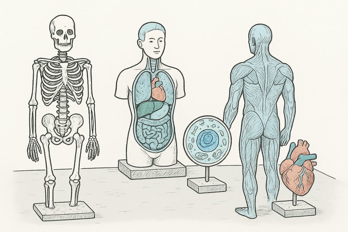
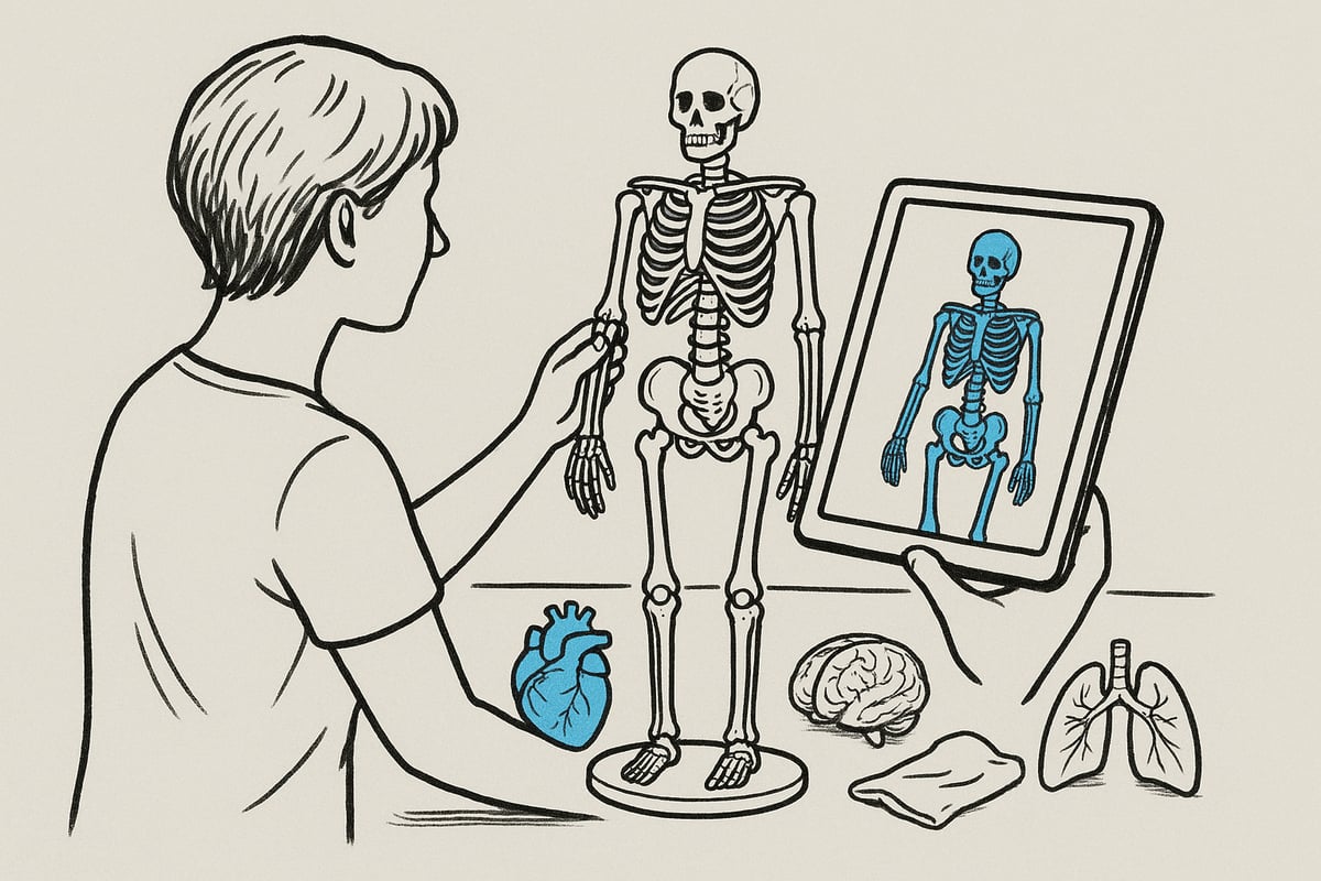
Skelet- og knoglemodeller
Skelet- og knoglemodeller er klassiske værktøjer i anatomiundervisning. De fås som fuldskala skeletter, enkelte knogler eller specifikke regioner som kranier og rygsøjler.
Disse modeller gør det let at demonstrere ledbevægelser, knoglestrukturer og biomekanik, hvilket styrker forståelsen af menneskets opbygning. Stan skeletmodellen er fx populær både i folkeskoler og på universiteter. Ifølge Optisafe er skeletmodeller nogle af de mest solgte, fordi en anatomi model af denne type gør det nemt at visualisere kroppens bevægeapparat.
De er uundværlige for fysioterapeuter og kiropraktorer, der skal forklare sammenhænge for patienter.
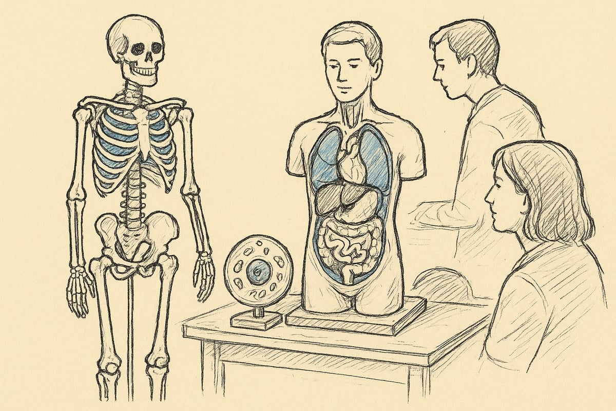
Organ- og torso modeller
Organ- og torso modeller viser kroppens indre organer som hjerte, hjerne, lunger og fordøjelsessystem. Disse modeller bruges til at forklare organfunktioner, sygdomme og behandlingsforløb på en håndgribelig måde. Ofte kan organerne tages ud og undersøges separat, hvilket giver en ekstra dimension til læringen.
Et eksempel er brugen af fordøjelsessystem-modeller i patientvejledning, hvor komplekse processer bliver letforståelige. Modellerne er centrale i både biologiundervisning og medicinstudier. Hvis du vil dykke dybere ned i mulighederne, kan du finde flere eksempler på anatomiske modeller af organer og strukturer, som bidrager til at øge forståelsen i sundhedsuddannelser.
En anatomi model af organer giver mulighed for at kombinere visuel og taktil læring, hvilket styrker både hukommelse og forståelse.
Muskelmodeller og bevægeapparat
Muskelmodeller illustrerer musklernes placering, opbygning og samspil med knogler og led. De bruges bredt i idræts- og fysiologiundervisning samt sportsmedicin, hvor det er vigtigt at kunne forklare, hvordan muskler arbejder sammen under bevægelse.
Ofte kombineres muskelmodeller med plakater og 3D-modeller for at give et komplet billede af bevægeapparatet. En anatomi model af muskler gør det muligt at demonstrere dynamiske processer, som ellers kan være svære at forklare abstrakt.
Disse modeller støtter både undervisere og studerende i at forstå komplekse biomekaniske sammenhænge.
Celle- og mikroskopimodeller
Celle- og mikroskopimodeller gør det muligt at visualisere strukturer, der normalt er usynlige for det blotte øje. Modeller af celler, virus og væv anvendes i gymnasier, universiteter og laboratorier til at undervise i mikrobiologi og genetik.
Ved at bruge en anatomi model på dette niveau kan man forklare processer som mitose, meiose og osmose på en måde, der engagerer både begyndere og avancerede studerende.
Disse modeller styrker forståelsen af livets grundlæggende byggesten og gør komplekse biologiske processer mere tilgængelige.
Specialmodeller: Patologiske tilstande og dyremodeller
Specialmodeller dækker alt fra sygdomsramte organer til dyreanatomi. Patologiske modeller viser fx kræft, åreforkalkning eller andre sygdomme, hvilket gør dem uundværlige i klinisk undervisning og patientinformation.
Veterinære anatomi modeller inkluderer hesteskeletter, dyreorganer og modeller til brug i veterinæruddannelser. En anatomi model af denne type hjælper med at formidle vigtige forskelle og ligheder mellem arter.
Disse specialmodeller bidrager til en mere nuanceret forståelse af både menneskets og dyrenes anatomi, hvilket styrker både forskning og praksis.
Sådan vælger du den rette anatomimodel i 2025
At vælge den rette anatomi model kan virke uoverskueligt med de mange muligheder i 2025. For at sikre det bedste læringsudbytte bør du først og fremmest afklare, hvad formålet med din anatomi model er. Skal den bruges til undervisning, patientvejledning, forskning eller måske til selvstudie? Hver anvendelse stiller forskellige krav til både detaljegrad og funktionalitet.
Overvej nøje, hvem der skal bruge modellen. Elever i folkeskolen har ofte brug for en mere simpel anatomi model, mens medicinstuderende eller sundhedsprofessionelle får mest ud af avancerede modeller med afmonterbare dele og tydelig farvekodning. Til patientvejledning kan en model med tydelige visuelle markeringer gøre komplekse diagnoser lettere at forstå.
Detaljeringsgraden spiller en central rolle. En simpel anatomi model kan dække basisbehov, men til specialiseret undervisning eller forskning bør du vælge en model med flere lag, adskillelige organer og realistisk farvesætning. Materialevalg har også betydning. Plastik og resin er populære for deres holdbarhed og lette rengøring, mens mere avancerede modeller kan fremstilles i naturmaterialer for øget realisme. Digitale modeller vinder også frem i takt med teknologiske fremskridt.
Størrelsen på din anatomi model skal matche både pladsforhold og anvendelse. Bordmodeller er praktiske til små hold eller selvstudie, mens fuldskala modeller egner sig til demonstrationer i større undervisningssammenhænge. Husk også at tænke på opbevaring og rengøring, især hvis modellen skal bruges ofte eller af mange.
Pris og budget spiller naturligvis ind. Kvalitet og funktionalitet bør altid sammenlignes, før du beslutter dig. Det kan være værdifuldt at indhente tilbud fra flere leverandører og undersøge garantier og supportmuligheder. Mange skoler og klinikker prioriterer modeller med lang holdbarhed, hvilket ofte retfærdiggør en højere investering.
Digital integration er en af de største trends i 2025. Flere og flere modeller er nu kompatible med AR og VR, hvilket åbner for nye muligheder i undervisning og fjernlæring. Ifølge Optisafe prioriterer 72% af sundhedsuddannelser i dag modeller, der kan kombineres med digitale løsninger. Interaktive modeller fra producenter som 3B Scientific gør det muligt at visualisere kroppens strukturer på helt nye måder.
Herunder ses en oversigt, der sammenligner klassiske og digitale modeller:
| Kriterium | Klassisk model | Interaktiv/digital model |
|---|---|---|
| Detaljeringsgrad | Høj/variabel | Meget høj, ofte 3D |
| Pris | Ofte lavere | Ofte højere |
| Vedligeholdelse | Let | Minimal |
| Læringsmuligheder | Taktil, visuel | Visuel, virtuel, AR/VR |
| Opdatering | Sjældent | Løbende via software |
Hvis du vil have yderligere vejledning om, hvordan du vælger og anvender en anatomi model som studerende, kan du finde praktiske tips og anbefalinger i Guide til studerende i anatomi.
Når du vælger din næste anatomi model, bør du altså tænke på både formål, målgruppe, detaljeringsgrad, materialer, størrelse, pris og digitale muligheder. På den måde sikrer du, at din investering både er fremtidssikret og giver maksimal læring og værdi.
Fremtidens trends inden for anatomimodeller
Fremtiden for anatomi model udvikling bringer markante forandringer. Nye teknologier, bæredygtige materialer og øget tilpasning sætter dagsordenen i 2025. Denne sektion giver dig indblik i de vigtigste trends, så du kan forstå, hvordan anatomi model løsninger udvikler sig og hvilke muligheder, der venter for undervisning, forskning og klinisk praksis.
Digitalisering og interaktive modeller
Digitaliseringen har transformeret anatomi model markedet. Virtuelle løsninger som AR og VR gør det muligt at udforske menneskekroppen i detaljer uden fysiske begrænsninger. Mange undervisere vælger digitale platforme, hvor elever kan dissekere og sammensætte organer på skærmen. Dette forbedrer forståelsen, især når det kombineres med fysiske modeller.
Eksempelvis er 3D-modeller, der styres via tablet eller mobil, blevet populære i fjernundervisning. Statistik viser, at brugen af digitale anatomi model løsninger er steget med 40 procent siden 2022. Udviklingen peger på endnu mere avancerede teknologier til simulering af kroppens strukturer.
Forskning i avanceret modelgenerering, som beskrevet i Steerable Anatomical Shape Synthesis, viser, hvordan neurale netværk og AI kan skabe endnu mere realistiske og dynamiske anatomi model oplevelser. Dette åbner for nye læringsmetoder og personaliserede simulationer.
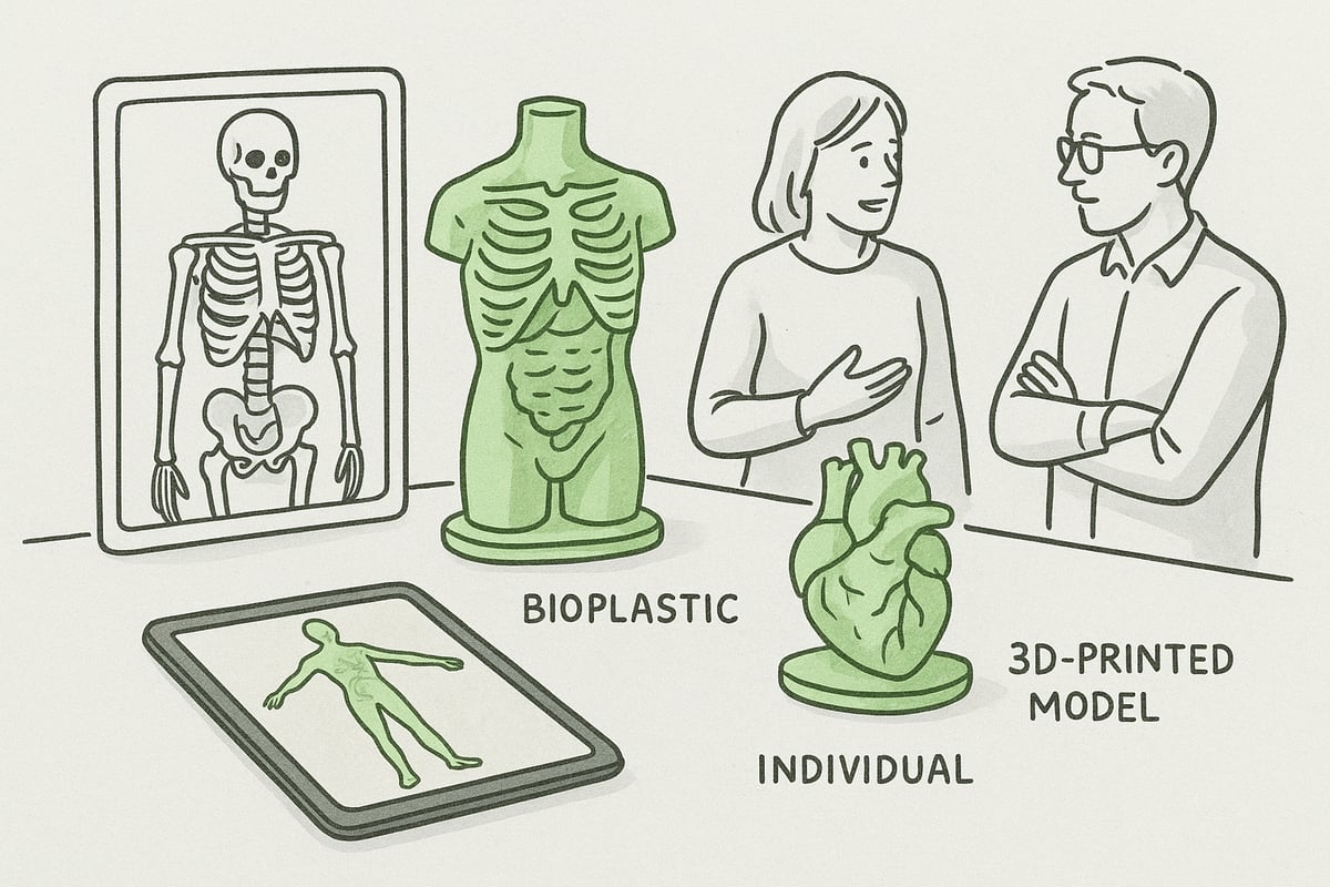
Bæredygtighed og materialevalg
Bæredygtighed er blevet et centralt emne ved valg af anatomi model. Mange producenter prioriterer nu miljøvenlige materialer såsom bioplast eller genanvendt plast for at mindske klimaaftrykket. Dette skift ses tydeligt i både skoler og sundhedsuddannelser, hvor der stilles krav om ansvarligt indkøb.
En anatomi model fremstillet af genanvendelige materialer har lang levetid og kan bruges igen og igen uden at gå på kompromis med kvaliteten. Derudover er produktionen ofte mere energieffektiv, hvilket bidrager til grøn omstilling i uddannelsessektoren.
For institutioner, der ønsker at kombinere innovation og miljøhensyn, er det muligt at vælge modeller, der både er holdbare og lette at rengøre. Dette gør dem velegnede til daglig brug og sikrer, at anatomi model løsninger følger tidens krav til bæredygtighed.
Individualisering og tilpasning
Efterspørgslen på skræddersyede anatomi model løsninger vokser hurtigt. Med 3D-print kan man nu fremstille modeller baseret på specifikke patientdata eller særlige undervisningsmål. Dette gør det muligt at illustrere sjældne sygdomme eller særlige anatomiske variationer, som ikke findes i standardmodeller.
Særligt i forskningsmiljøer og specialuddannelser har individualiserede modeller stor værdi. En anatomi model kan tilpasses både i størrelse, farvekodning og detaljeringsgrad, så den matcher præcis det behov, brugeren har. Det giver større fleksibilitet og gør undervisningen mere relevant.
Tilpasning betyder også, at modeller kan opdateres løbende i takt med ny viden. Det sikrer, at undervisningsmaterialet altid er aktuelt, og at elever og professionelle får den mest korrekte information i deres anatomi model arbejde.
Globalisering og tilgængelighed
Globaliseringen har gjort det nemmere end nogensinde at få adgang til den rette anatomi model. Flere leverandører tilbyder nu internationale løsninger med support på mange sprog. Dette gør det let for både danske og udenlandske institutioner at bestille og tilpasse modeller online.
Online platforme har øget tilgængeligheden, så man hurtigt kan sammenligne priser, materialer og funktionalitet. Statistik viser, at 60 procent af danske institutioner nu køber deres anatomi model online, hvilket sparer tid og giver større udvalg.
Denne udvikling understøtter den fortsatte professionalisering af undervisning og patientkommunikation. Uanset om du er underviser, studerende eller professionel, har du adgang til de bedste og mest opdaterede anatomi model løsninger fra hele verden.
Praktisk brug: Maksimer læringsudbyttet af anatomimodeller
At arbejde med en anatomi model i praksis åbner for en dybere forståelse af kroppens opbygning. Uanset om du underviser, træner, eller studerer selv, kan den rette tilgang gøre forskellen på overfladisk viden og ægte indsigt. Her får du konkrete strategier til at maksimere udbyttet af din anatomi model i hverdagen.
Didaktiske strategier i undervisning
Når du integrerer en anatomi model i undervisningen, styrker du både det taktile og det visuelle element. Hands-on aktiviteter, hvor elever selv samler eller undersøger modeller, motiverer til aktiv deltagelse og forbedrer forståelsen.
Det er effektivt at kombinere modeller med plakater og digitale ressourcer. For eksempel kan en skeletmodel suppleres med anatomiske plakater designet i Danmark for en mere nuanceret tilgang. Case-baseret læring, hvor elever løser opgaver ud fra modeller, træner desuden både analyse og samarbejde.
En anatomi model gør teori konkret. Elever får mulighed for at undersøge led, muskler og organer tæt på, hvilket giver dem indsigt, der ofte overstiger traditionel tavleundervisning.
Patientkommunikation og formidling
En anatomi model er et stærkt værktøj i patientkommunikation. Læger og sundhedspersonale bruger ofte modeller til at forklare diagnoser, behandlinger eller kirurgiske indgreb. Dette bidrager til at øge patientens forståelse og tryghed, især når komplekse sammenhænge skal formidles.
Ved at pege på et specifikt område på en model kan du illustrere, hvor og hvordan en skade, operation eller sygdom påvirker kroppen. Mange patienter oplever, at en visuel forklaring gør det lettere at stille spørgsmål og engagere sig i deres egen behandling.
Modeller af hjerte, led eller organer er særligt populære, fordi de kan skilles ad og vise indre strukturer. For sundhedspersonale, der ønsker at styrke deres formidling, anbefales det at udforske ressourcer til undervisere i anatomi for inspiration og vejledning. En anatomi model i konsultationen kan gøre forskellen på forvirring og klarhed for patienten.
Selvstudie og eksamensforberedelse
For studerende, der selv arbejder med en anatomi model, er fordelene markante. Fysiske modeller gør det muligt at repetere og visualisere komplekse anatomiske sammenhænge, hvilket styrker både forståelse og hukommelse.
En undersøgelse fra Scandidact viser, at 85% af medicinstuderende foretrækker fysiske modeller fremfor udelukkende digitale løsninger. Dette skyldes blandt andet, at en anatomi model engagerer flere sanser og gør det lettere at huske detaljer.
Selvstudie med modeller kan kombineres med quizzer, flashcards eller digitale platforme, så læringen bliver varieret og effektiv. Ved at bruge modellen aktivt, frem for blot at læse, får du en mere robust og vedvarende viden.
Vedligeholdelse og opbevaring af modeller
For at sikre lang levetid og optimal funktion er korrekt vedligeholdelse af din anatomi model afgørende. Rengør modellen jævnligt med egnede midler, og undgå udsættelse for direkte sollys og fugt, da det kan skade materialet.
Opbevar modellerne i stabile opbevaringskasser, gerne mærket for let adgang og organisering. På skoler og klinikker er det en fordel at have faste rutiner for rengøring og kontrol, så modellerne altid fremstår pæne og klar til brug.
En god anatomi model kræver også løbende opdatering. Overvej at supplere med nye modeller eller reservedele, når medicinsk viden udvikler sig, eller hvis slid og brug kræver det. På den måde sikrer du, at både lærere og studerende får det bedste udbytte af deres investering.
Guide: Trin-for-trin til valg og brug af anatomimodel
At vælge den rette anatomi model kræver overblik og systematik. Følg denne guide trin for trin, så du får mest muligt ud af din investering og sikrer optimal læring, uanset om du er underviser, studerende eller professionel.
Trin 1: Identificér dit behov
Start med at definere præcist, hvad din anatomi model skal bruges til. Skal den understøtte undervisning, patientvejledning, forskning eller måske et personligt projekt? Overvej også målgruppen. Er det begyndere, avancerede brugere eller specialister? En tydelig behovsafklaring sikrer, at du vælger en model, som matcher dine læringsmål og det faglige niveau. Tænk også over, om modellen skal bruges ofte eller kun lejlighedsvis. Det sparer både tid og ressourcer på sigt.
Trin 2: Vælg modeltype og detaljeringsgrad
Dernæst vælger du den type anatomi model, der bedst matcher dit formål. Skal det være et skelet, organ, muskelmodel eller måske en cellemodel? Vurder, om det er vigtigt, at modellen kan skilles ad, har farvekoder eller er interaktiv. Nogle modeller egner sig bedst til at demonstrere bevægelser, mens andre giver et detaljeret indblik i indre strukturer. Jo mere præcis du er i dit valg, desto større udbytte får du af modellen i undervisningssammenhænge.
Trin 3: Undersøg materialer og kvalitet
Materialevalg har stor betydning for både holdbarhed og realisme. En anatomi model i plastik er ofte let og robust, mens resin giver større detaljerigdom. Overvej også om bæredygtighed er vigtig for dig – flere producenter tilbyder miljøvenlige alternativer. Tjek, hvordan modellen skal rengøres og vedligeholdes, så den forbliver hygiejnisk ved hyppig brug. Kvaliteten skal matche dine krav til både udseende og funktionalitet.
Trin 4: Sammenlign priser og leverandører
Indhent tilbud fra flere leverandører, og sammenlign priser i forhold til kvalitet, garanti og support. Det er en fordel at vælge en leverandør med dokumenteret ekspertise og bredt sortiment. For eksempel kan du læse mere om kvalitet og service hos Om eAnatomi og vores ekspertise, som er kendt for dansk design og faglighed. Husk at undersøge leveringsbetingelser og eventuelle muligheder for tilpasning.
Trin 5: Integrér modellen i undervisning eller praksis
Planlæg, hvordan anatomi model skal bruges i praksis. Skal den indgå i demonstrationer, gruppearbejde, eller patientvejledning? Kombinér fysiske modeller med digitale ressourcer for at skabe dybere forståelse. Udnyt modellens muligheder for at engagere brugerne aktivt – det styrker både indlæring og motivation. Brug også modellerne til at illustrere komplekse sammenhænge, som kan være svære at forklare med ord alene.
Trin 6: Vedligehold og opdater løbende
For at sikre lang levetid skal du rengøre og opbevare anatomi model korrekt. Brug milde rengøringsmidler og følg producentens vejledning. Overvej at opbevare modellerne i kasser eller skabe med tydelig mærkning, så de er lette at finde og beskytte mod skader. Hold dig opdateret med nye modeller og opdateringer, så undervisningen altid er fagligt tidssvarende. Det giver både sikkerhed og kvalitet i det daglige arbejde.
Når du nu har fået et overblik over de mange muligheder og fordele ved anatomimodeller, er du allerede godt på vej til at styrke din forståelse af kroppens kompleksitet – uanset om du underviser, studerer eller arbejder professionelt med sundhed. Vi har gennemgået både klassiske og digitale løsninger, materialer, og hvordan modeller kan gøre læring mere levende og effektiv. Hvis du vil dykke dybere ned i udvalget eller finde den model, der matcher netop dine behov i 2025, kan du læse mere og få inspiration her: Læs mere her
0 comments