
Did you know that original anatomical illustrations still play a vital role in medicine, education, and art, even in a digital age? This guide takes you on a journey through their fascinating history, contemporary uses, and future trends through 2025.
You will gain insight into how original anatomical illustrations differ from digital or mass-produced alternatives, and why they continue to have unique value. The article will equip you to choose quality illustrations and understand their importance.
Be inspired by the possibilities and take the first step with our guide to selecting and applying these unique works in practice.
The Story Behind Original Anatomical Illustrations
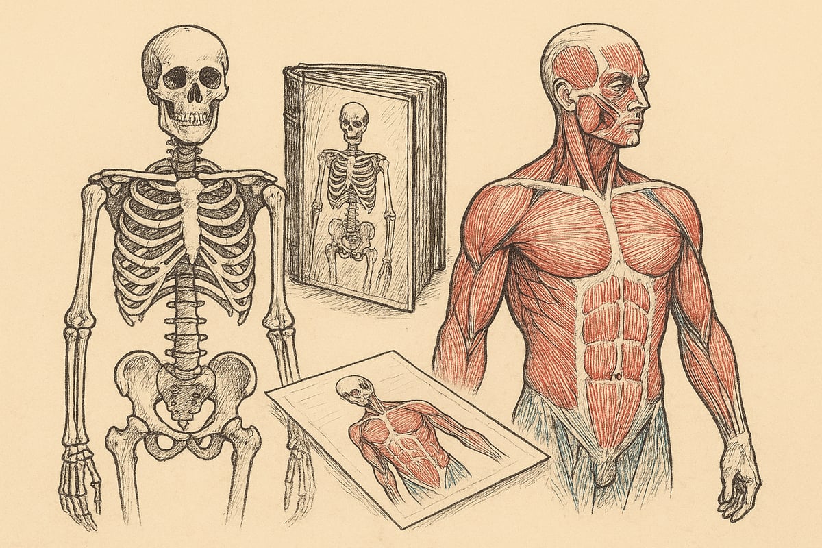
Original anatomical illustrations have played a central role in the understanding of the human body throughout history. These works combine artistic craftsmanship and scientific accuracy, making them indispensable in both medicine and art. The development of these illustrations reflects not only technological advances, but also changing views of the body and knowledge.
The development from classic to modern illustrations
For centuries, original anatomical illustrations have been essential to medical knowledge. In the Renaissance, Leonardo da Vinci revolutionized the field with his detailed sketches, which set new standards for both accuracy and aesthetics. Since then, hand-drawn illustrations have been the foundation of textbooks and scientific publications.
Examples such as Vesalius' "De humani corporis fabrica" from 1543 demonstrate how art and science went hand in hand. Statistics show that the number of anatomical illustrations in textbooks grew sharply from the 16th century to the present day, when almost all medical works contain visual explanations. Classical techniques still inspire today's artists, who often mix tradition with modern expressions to create unique works.
Famous artists and their contributions
Several pioneers have had a major impact on the development of original anatomical illustrations. Andreas Vesalius was one of the first to combine precise observation and artistic representation, which still inspires today. Modern Danish and international illustrators continue to build on this legacy, often in close collaboration with doctors.
The collaboration between artists and medical experts has resulted in award-winning illustrations used in both research and teaching. For an in-depth look at Vesalius' work, read more about Andreas Vesalius' Contributions to Anatomy . These works not only have scientific value, but have also left a lasting mark on art history.
The transition to digital and hybrid techniques
The introduction of digital tools has changed the way original anatomical illustrations are created. Digitization allows for faster and more precise work, but many still prefer hand-drawn works for their authenticity and unique expression. Statistics show that digital illustrations now make up the majority of teaching materials, but the demand for original illustrations remains high.
The use of 3D modeling and augmented reality has further expanded the possibilities for communication and learning. Yet many artists choose to combine traditional and digital techniques, resulting in hybrid illustrations that bring together the best of both worlds.
Collectibles and cultural significance
Original anatomical illustrations are now considered valuable collectors' items and investments. They often adorn museums and public exhibitions, conveying both scientific and aesthetic value. For art collectors and professionals, these works have a special status, as they represent both historical insight and artistic skill.
Many collectors seek out rare illustrations from famous periods or artists. The market for these works is growing, and they are often used as reference points for contemporary artists. Their cultural significance cannot be overstated, as they continue to inspire and educate new generations.
Uses for Original Anatomical Illustrations
Original anatomical illustrations have a surprisingly wide range of uses, from teaching to art and collectibles, adding depth, authenticity and aesthetic value to any subject area where understanding anatomy is central.
Education and research
In the education sector, original anatomical illustrations play a crucial role in promoting understanding of complex biological structures. Medical students and educators use illustrations to visualize organs, muscles, and bones, significantly increasing learning outcomes.
Studies show that students experience better retention of knowledge when they have access to visual aids. Illustrations make it easier to explain difficult concepts and create a concrete connection between theory and practice. Many educators choose anatomical illustrations for teaching to ensure high quality and pedagogical value.
In addition, original anatomical illustrations are often used in research articles and presentations, where precision and detail are essential for communicating new discoveries.
Clinical practice and patient communication
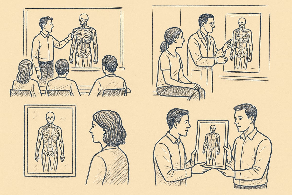
In clinical practice, original anatomical illustrations help doctors and nurses explain diagnoses and treatment options to patients. A well-executed illustration can make complex medical concepts more accessible and understandable to everyone.
Patient brochures with original anatomical illustrations increase patient understanding and compliance. When patients see an accurate representation of their condition, they feel more confident and informed. This strengthens the dialogue between the practitioner and the patient, which can result in better treatment outcomes.
Furthermore, illustrations are used in clinical conferences and interdisciplinary meetings, where they facilitate communication between specialists.
Art and design
Original anatomical illustrations inspire artists and designers worldwide. In modern art, they are used as a starting point to explore the shapes and proportions of the human body, while in graphic design they add both aesthetics and authenticity.
Many designers incorporate original anatomical illustrations into fashion, posters and interiors, creating a unique visual expression. Collaborations between artists and medical experts create groundbreaking works where science and art merge.
This interdisciplinary approach opens up new interpretations and innovative expressions, where classic techniques meet modern trends.
Public information and exhibitions
Museums and exhibition spaces use original anatomical illustrations to convey knowledge about the structure of the body to a wide audience. These illustrations make complex topics easy to understand and engaging, increasing interest in science.
Successful exhibitions in Scandinavia have attracted many visitors, partly due to the fascinating original anatomical illustrations presented. Statistics show increasing visitor numbers at exhibitions where illustrations are a central element.
Furthermore, illustrations are included in public information campaigns, where they are used to explain health topics in an easy-to-understand way.
Collectibles and gift items
The market for original anatomical illustrations as collectibles and gifts is growing steadily. Many collectors value the unique works for their artistic and historical value, while professionals often receive them as exclusive gifts.
Limited edition prints and signature works are particularly sought after, especially when they combine classic craftsmanship with modern techniques. In-demand motifs include both human anatomy and rare biological structures.
The trend indicates that original anatomical illustrations are not only useful, but also valued investments and personal gifts.
How to Choose Quality Anatomical Illustrations
Finding quality original anatomical illustrations requires insight into authenticity, materials, style, price, and purchasing options. Each field has its own evaluation criteria that ensure both aesthetic and scientific value. This guide will help you make informed choices, whether you are a collector, educator, or art enthusiast.
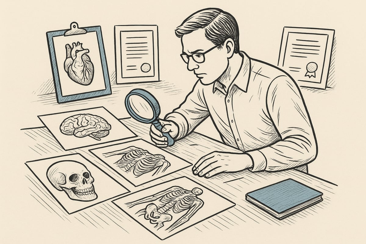
Criteria for authenticity and originality
When evaluating original anatomical illustrations, authenticity should always be your top priority. Look for documentation, such as certificates, signatures, or provenance, that confirms the work's origin. Genuine illustrations often have unique characteristics such as hand-drawn lines, paper patina, and personal touches from the artist.
Fake illustrations can often be identified by missing details, machine printing, or the absence of accompanying documents. Compare the work to known examples to ensure it is distinct from mass-produced copies.
A short checklist:
- Certificate of Authenticity
- Visible signature or stamp
- Documented provenance
By prioritizing these elements, you can ensure that your original anatomical illustrations are both authentic and valuable.
Materials and printing techniques
The choice of materials plays a major role in the durability of original anatomical illustrations. Many artists use archival-quality paper, canvas, or specialized techniques such as watercolor and ink. Each material provides a unique texture and aesthetic that can enhance the value of the work.
Digital prints can offer beautiful reproductions, but often lack the depth and texture that traditional materials provide. Statistics show that collectors and educators prefer hand-drawn works on acid-free paper, as they retain detail and color best over time.
Table: Materials and their advantages
| Material | Advantage | Durability |
|---|---|---|
| Paper (archive) | Natural texture | High |
| Canvas | Robust and classic | Very high |
| Digital printing | Consistent quality | Varies |
When choosing original anatomical illustrations, you should always inquire about the quality of the materials.
Style and level of detail
The style of original anatomical illustrations ranges from hyper-detailed, scientific renderings to more stylized, artistic interpretations. Both styles have their place, depending on the purpose. For medical education, scientific accuracy is highly valued, while art collectors may prefer unique styles and creative expressions.
A good example of Danish design tradition and style can be found among anatomical posters designed in Denmark , where the balance between aesthetics and professionalism is clear.
Always consider whether the level of detail in the illustration matches your needs. Is there a focus on precise structures, or is there room for interpretation? By choosing the right style, you ensure that your original anatomical illustrations meet both practical and aesthetic requirements.
Pricing and investment
The price of original anatomical illustrations depends on several factors: the artist's reputation, the age of the work, the materials and rarity. Rare, signed illustrations from well-known artists often sell for significantly higher prices than newer or mass-produced works.
The market for these illustrations has seen increasing interest from both investors and collectors. Consider the potential appreciation of the work and research past price trends before making a purchase.
Good advice for investing:
- Choose works with documented provenance
- Research market trends
- Focus on reputable artists
By following this advice, you can get both pleasure and financial benefit from your original anatomical illustrations.
Buying and ordering illustrations
When you're ready to buy original anatomical illustrations, you should start by researching reliable artists and dealers. Visit galleries, online platforms, and specialized webshops where you can see examples and read reviews.
When ordering custom illustrations, it is important to communicate your wishes clearly and ensure that the artist has experience with anatomical motifs. Always ask for previous work and confirm delivery times.
Tips for buying and ordering:
- Examine the artist's portfolio
- Agree on price and delivery terms in advance
- Ask for documentation of authenticity
By following these steps, you will have greater confidence that your original anatomical illustrations will meet your expectations.
Modern Trends and the Future of Anatomical Illustrations
The development of original anatomical illustrations is going strong these years. New technologies, sustainability, personalization and interdisciplinary collaborations are shaping the future of the field. In this section, you will get an overview of the most important trends that will characterize the area in 2025.
Technological advances and digital integration
Technology has revolutionized how original anatomical illustrations are created and used. 3D modeling, virtual reality and augmented reality make it possible to visualize the body's structures in completely new ways. Statistics show a significant increase in the use of digital solutions globally, especially in the education sector. Yet hand-drawn illustrations are experiencing a renaissance, as many appreciate their unique and authentic expression. The combination of classic and digital methods creates exciting hybrid solutions that inspire both educators and artists. If you want to delve deeper into the development, you can read more about The History of Anatomical Illustrations .
Sustainability and ethical considerations
Sustainability is taking on more importance in the production of original anatomical illustrations. Many artists are choosing environmentally friendly materials such as recycled paper and non-toxic paints. At the same time, there is a focus on ethical guidelines, for example in relation to the use of medical data and correct representation of anatomy. Responsible initiatives promote transparency and quality in the industry, which strengthens trust among both collectors and educational institutions. This development contributes to the illustrations not only being beautiful, but also being produced with consideration for the environment and ethics.
Personalization and user engagement
A clear trend is the demand for personal and customized original anatomical illustrations. Several artists now offer to adapt motifs to specific requests, e.g. for teaching, research or collectors. Statistics show a growing interest in personalized solutions, where the user is actively involved in the process. This can both enhance the learning outcome and make the works unique. If you want to order a personal illustration, you can follow this Guide to ordering illustrations .
Interdisciplinary collaborations and globalization
Original anatomical illustrations are increasingly being created through collaborations between doctors, artists and technologists. International projects and exchange programs create new opportunities for innovation and knowledge sharing. Global trends show that the demand for illustrations is increasing, not only in classic textbooks, but also in digital platforms and exhibitions. This development emphasizes the importance of interdisciplinarity and cultural exchange when developing the illustrations of the future.
Future challenges and opportunities
The future of original anatomical illustrations holds both challenges and great opportunities. Copyright and digital copying require clear guidelines to protect artists and maintain quality. At the same time, technological advances are opening up new applications, such as interactive learning tools and integrated AR solutions. Market forecasts point to continued growth through 2025, especially among educators, collectors and designers. For those looking to invest in the area, it is important to keep up with both technological and artistic trends.
Guide: Step-by-Step To Creating Your Own Original Anatomical Illustration
Creating original anatomical illustrations requires both accurate professional references and creativity. This guide walks you through five essential steps to help you develop your own work, whether you're a student, teacher, or artist.
Step 1: Research and reference collection
The first step towards creating original anatomical illustrations is thorough research. Start by finding reliable sources, such as scientific articles, museums, and reputable textbooks. A popular reference is the Musculoskeletal Anatomy Textbook , which offers detailed illustrations and explanations.
Collect images, diagrams, and descriptions of the anatomical area you wish to illustrate. Use databases and museum digital archives to ensure accuracy. Compare multiple sources to avoid errors in your work.
Make a list of the most important structures and variations. This will help you understand the complexity and focus on the most important details when you start drawing your own original anatomical illustrations.
Step 2: Sketching and composition
Once you have gathered your reference base, the next step is to develop sketches and compositions. Start with simple shapes to establish proportions and placement. Sketching is essential for achieving balance in original anatomical illustrations.
Draw several variations to experiment with perspective and structure. Use light pencil strokes so you can easily correct mistakes along the way. Think about how to emphasize elements and where to place text explanations.
Consider creating small thumbnails or partial sketches before moving on to the final layout. Good composition makes your original anatomical illustrations both informative and visually appealing.
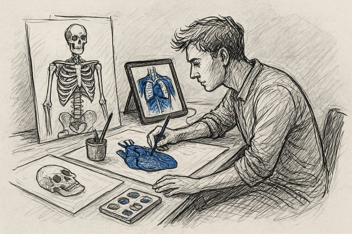
Step 3: Execution with selected materials
Now it's time to choose materials and begin the actual drawing. Many choose pencil, ink, or watercolor, but more also use digital tools, depending on the purpose of their original anatomical illustrations.
Choose archival-quality paper to ensure durability if you work in analog. Digital illustrations require high resolution and color calibration to achieve sharp lines and precise shades.
Test different materials to find the style that best supports your message. Remember that material choice can affect the expression and readability of original anatomical illustrations, so be careful at this stage.
Step 4: Detail and scientific precision
Once the basic shape is in place, focus on detail and accuracy. Add anatomical details, such as nerves, blood vessels, and muscle structures, to make original anatomical illustrations both accurate and educational.
Collaborate with professionals, such as doctors or biologists, to validate your work. Use clear text explanations and markings so that the illustrations can be understood without prior expertise.
Be aware of common errors, such as incorrect proportions or missing structures. The quality of original anatomical illustrations depends on the balance between scientific precision and aesthetic presentation.
Step 5: Final presentation and sharing
Finally, your original anatomical illustrations need to be presented professionally. Scan or photograph the work in high resolution, and easily edit the image for optimal sharpness and color reproduction.
Consider framing or digital presentation depending on purpose. Share the illustrations on social media, educational networks, or at exhibitions to gain visibility.
Through a careful process, you ensure that your original anatomical illustrations both convey knowledge and appear as unique works of art.
When we look at how original anatomical illustrations have shaped both medical knowledge and creative inspiration over time, it becomes clear how valuable they are for both teaching, research and collectors. In this guide, you will gain insight into the history, techniques and many uses – as well as concrete advice on choosing illustrations that match your needs. If you are curious to go even deeper into quality illustrations, materials and the latest trends, you can delve further into the subject here: Read more here
0 comments