Forstørret pædagogisk hjerne, 4 dele - eAnatomi

Gehirn, vergrößertes pädagogisches Anatomiemodell, 4 Teile

€249,95
Zu Produktinformationen springen
Forstørret pædagogisk hjerne, 4 dele - eAnatomi
1/9

Gehirn, vergrößertes pädagogisches Anatomiemodell, 4 Teile

SKU: A614
€249,95
Inkl. Steuern. Versandkosten werden beim Checkout berechnet.
Auf Lager

Shop-Informationen anzeigen

Gehirn, vergrößertes pädagogisches Anatomiemodell, 4 Teile

Beschreibung

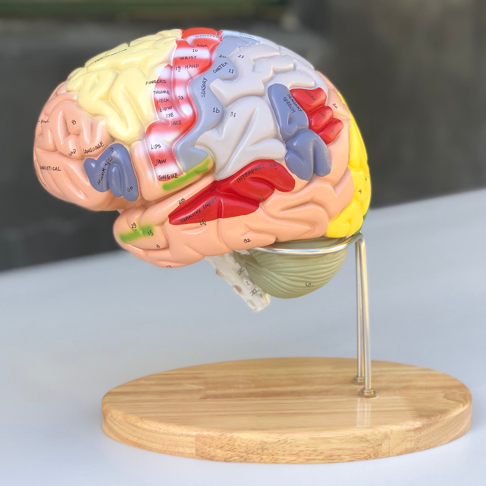
Wenn Sie ein vergrößertes, inspirierendes und äußerst didaktisches Gehirnmodell suchen, mit dem sich wichtige Bereiche des Gehirns so einfach wie möglich identifizieren lassen, empfehlen wir dieses wärmstens.


Das Modell ist aus sehr hartem und robustem Kunststoff gegossen. Im Gegensatz zu vielen unserer anderen Gehirnmodelle, die aus hohlem und flexiblem Kunststoff gegossen sind, lässt sich das Material dieses Modells weder zusammendrücken noch bewegen. Manche werden meinen, dass es dadurch weniger angenehm zu berühren und zu bearbeiten ist, wenn es auseinandergenommen und studiert werden soll. Andere halten dies für einen Vorteil.


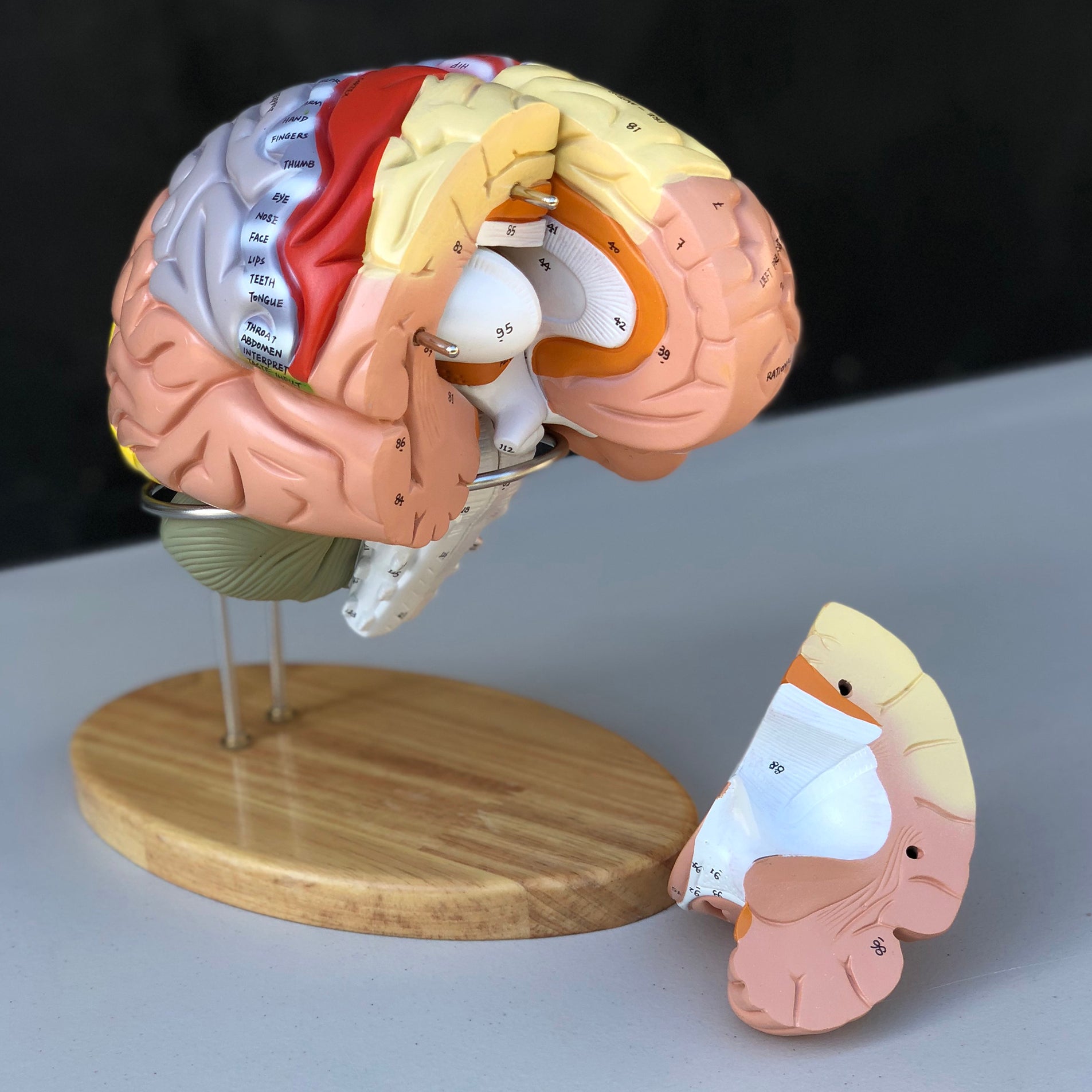
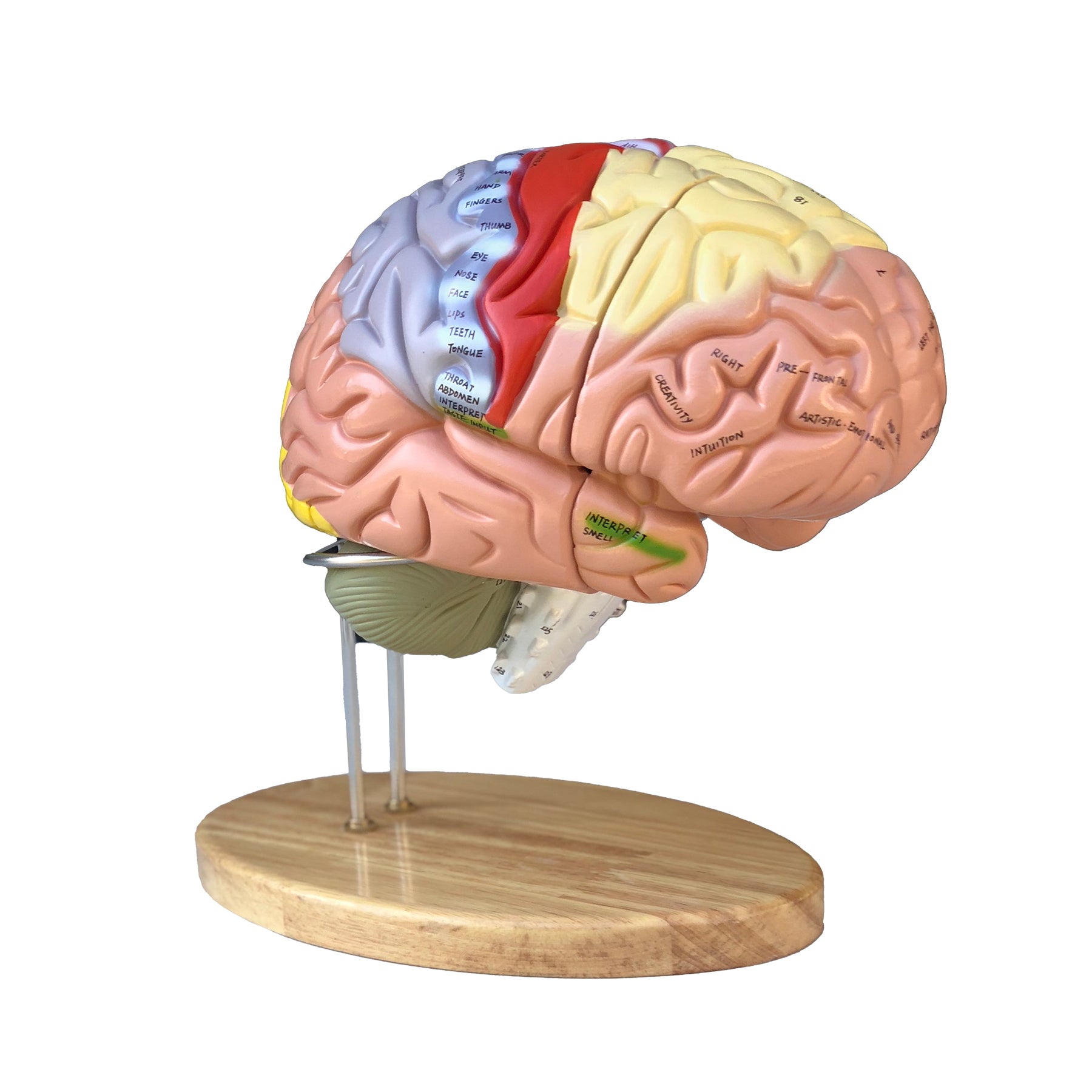
Das Modell kann in 4 Teile zerlegt werden, die durch Metallstifte zusammengehalten werden. Nachfolgend können Sie mehr über die anatomischen Details erfahren, wie z. B. die farbigen Bereiche und das limbische System. Im Vergleich zu einer erwachsenen Person ist das Modell etwa 1,5-mal normal groß. Die Abmessungen betragen 22 x 18,5 x 19 cm, und das Gewicht beträgt 1,9 kg. Das Modell ruht auf einem Metallständer, der auf einer schönen Basis aus lackiertem Eichenholz mit abgerundeten Kanten montiert ist (siehe die Bilder links). Das Modell lässt sich sehr einfach abnehmen und zerlegen.


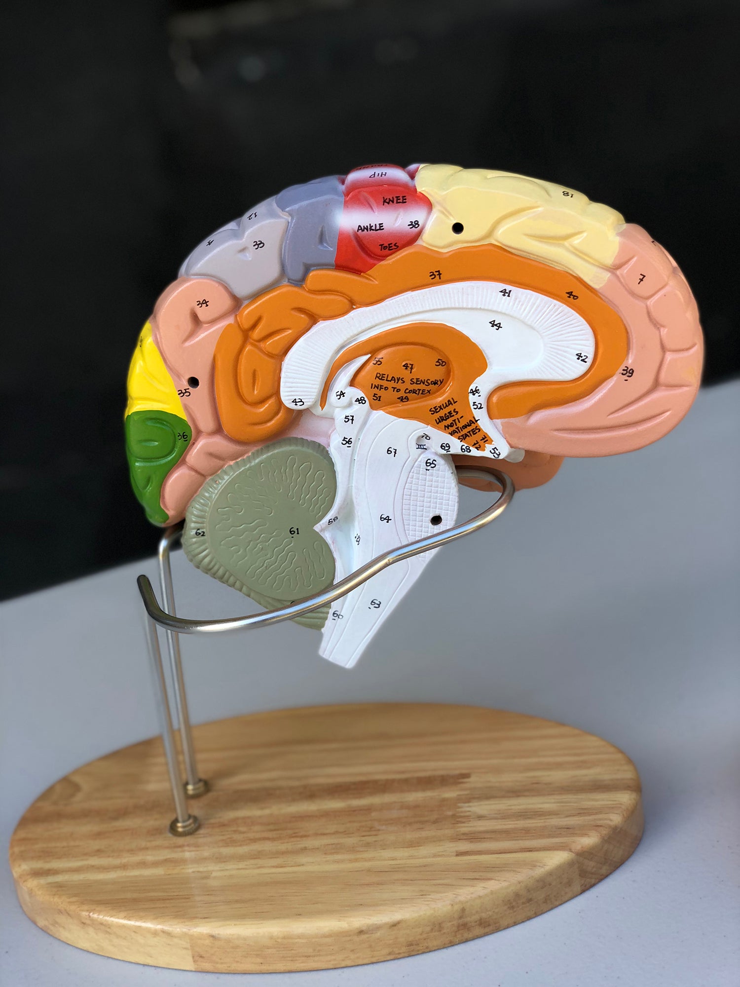
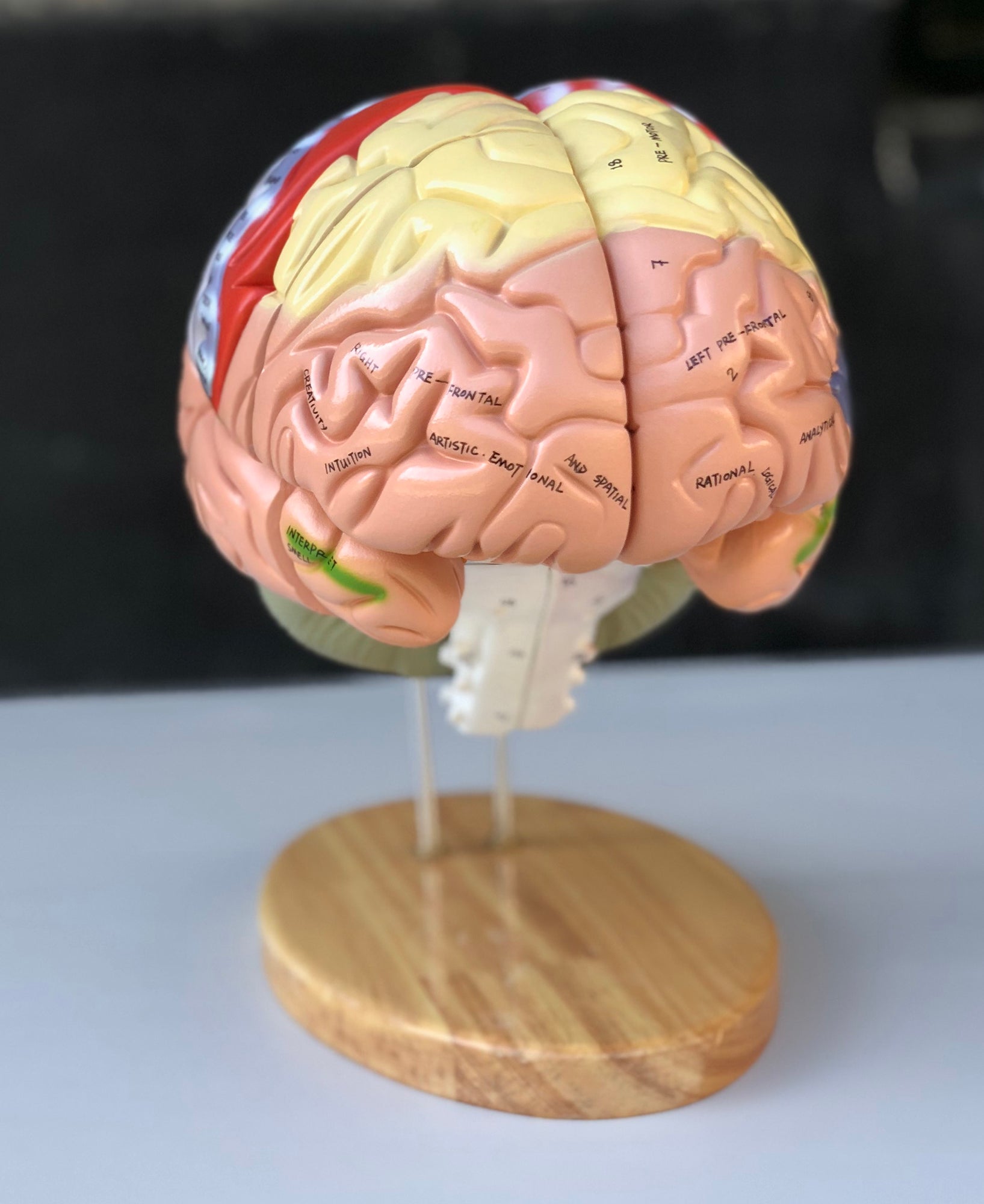
Es ist hervorzuheben, dass dieses Gehirnmodell das einzige in unserem Sortiment ist, das sowohl mit Nummerierungen wichtiger anatomischer Strukturen als auch mit direkt auf spezielle Bereiche geschriebenen Wörtern geliefert wird, wie z. B. dem primären motorischen Bereich (siehe die Bilder links und lesen Sie unten mehr).


NB: Die Nummerierungen und Bezeichnungen dienen als Orientierung. Gehen Sie daher bei der Verwendung kritisch vor, da sich eine Zahl bei einer anatomischen Struktur beispielsweise an der Grenze zu einer anderen Struktur befinden kann.

Anatomisch gesehen

Anatomically, the model shows the human brain, which can generally be divided into the cerebrum (cerebrum), the cerebellum (cerebellum) and the brain stem (truncus encephali).


These 3 structures are clearly separated via different colors, but the difference between gray and white matter cannot be seen on this model.


In the cerebrum (telencephalon and diencephalon), the lobes of the brain, as well as the thalamus and hypothalamus (and the pituitary gland) are primarily seen


In the cerebellum, the vermis cerebelli and the cerebellar hemispheres (hemisperium cerebelli) are seen


The brainstem shows its 3 parts (the midbrain, the pons and the medulla oblongata) as well as the apparent origins of the cranial nerves using Roman numerals (also called the cranial nerves)


Other structures such as the brain stem, fornix, ventricular system and the first 2 cranial nerves (the olfactory and optic nerves) are also seen, which do not originate from the brainstem


The pictures on the left show how the model can be separated into 4 parts. This makes it possible to study the internal structures of the brain, and some of these are seen in 3 dimensions.


Colored areas


The colors on this model are muted and very different. As seen in the images on the left, it is not only the lobes of the brain that are colored. Colors are also seen in special areas such as the pre- and postcentral gyrus, Broca's area, Wernicke's area and deeper structures.


As the model is both educationally colored and comes with English words written directly on the important areas of the model, you can very easily and quickly identify areas of interest. Examples are the primary motor area (gyrus precentralis) with indication of body regions ("neck, hip, shoulder, fingers" etc.) and areas such as "language" and speech".


The limbic system


Many of our customers ask about the limbic system in connection with the purchase of brain models. Hence this description.


The limbic system includes various anatomical structures in the central nervous system (CNS), and is primarily responsible for emotional functions such as anxiety, aggressiveness, mood, memory and social adaptability. Clinically, it is therefore often related to psychiatric disorders.


The limbic system includes, among other things amygdala, hippocampus, gyrus parahippocampalis, hypothalamus, fornix, corpus mammillare, the prefrontal cerebral cortex and the monoaminergic systems of the brainstem. The list is quite a bit longer - especially because numerous fiber connections connect the limbic structures. Many customers ask in particular about the amygdala and hippocampus (which is why they are mentioned first in this section).


NB: In this brain model, the hippocampus can be seen as well as some of the other limbic structures such as the fornix - but not the amygdala.


The amygdala is involved in anxiety and emotional coloring of sensory impressions. It lies as an almond-shaped nucleus IN FRONT of the hippocampus in the anterior pole of the temporal lobe (amygdala and hippocampus are therefore separate).


The hippocampus is involved in memory. It lies as an irregular twisted structure in the medial part of the temporal lobe.


If one of these 2 structures is to be seen clearly on a brain model, the brain tissue must be shown in a so-called frontal/coronal section through the temporal lobe. The frontal incision roughly corresponds to the incision direction "from ear to ear".


Because the amygdala is IN FRONT of the hippocampus (roughly speaking further forward "toward the forehead"), both of these structures can only be seen on a brain model if the model includes at least 2 frontal sections through the temporal lobe - or if the brain model is partially transparent.


We have not yet seen a brain model that shows 2 cuts through the temporal lobe, so that both the amygdala and the hippocampus are seen. In our range, on the other hand, we have a partially see-through brain model in the highest price range, which shows both structures.


All brain models in our range can be separated into different parts. All models (both with and without educational colors) that can be separated into 4 or more parts show the hippocampus. On almost all of these models, the hippocampus is also numbered and named on an overview that can be downloaded from the product descriptions of the brain models.

Bewegungstechnisch
Klinisches Set

Aus klinischer Sicht ist dieses Modell unser didaktisch wertvollstes, wenn ein Gehirnmodell zum Verständnis von Störungen (Symptomen) und Schäden verwendet werden soll, die durch Läsionen in Bereichen der Großhirnlappen verursacht werden. Diese Störungen und Schäden können beispielsweise Beeinträchtigungen der Bewegungen, des Tastsinns, der Sprache und des Sehvermögens sein.


Das Modell kann auch zum Verständnis vieler anderer Läsionen und Erkrankungen wie z. B. Epilepsie, Hirntumoren, Hydrozephalus und Läsionen verwendet werden, die Hirnnerven betreffen.


Obwohl die Durchblutung des Gehirns am Modell nicht sichtbar ist, kann es auch zum Verständnis von Apoplexien (Schlaganfällen) verwendet werden.

KUNDENSERVICE

Bestellwaren

Wenn das Produkt als Bestellung gekennzeichnet ist, bedeutet dies, dass das Produkt eine Größenordnung oder ein so hohes Qualitätsniveau hat, dass es nur auf Bestellung beschafft wird. Die Lieferzeit kann variieren, aber der Preis bleibt immer derselbe! Kontaktieren Sie uns für weitere Informationen, wenn Sie bestellen möchten.

Preise und Zahlungsmethoden
  • Die Preise sind in DKK inkl. MwSt. angegeben.
  • Du kannst mit folgendem bezahlen:
    - EAN-Nr.
    - MobilePay
    - Visa, Mastercard
Widerrufsrecht & Rückgabe
  • Widerrufsrecht besteht 14 Tage ab Lieferung.
  • Rücksendekosten betragen 60,- bei Kauf des Rücksendelabels bei uns (wird als PDF-Datei per E-Mail versandt) oder 0,- bei persönlicher Rücksendung an unsere Adresse.
  • Sehen Sie sich unsere Verkaufsbedingungen hier an.
Lieferung / Abholung

Wenn der Artikel als auf Lager angegeben ist, befindet er sich physisch an unserer Adresse in Frederiksberg und kann daher versandt oder abgeholt werden. Wählen Sie bei der Bestellung, was Sie wünschen.

Wenn Sie den Artikel abholen möchten, kann dies oft unmittelbar nach der Bestellung erfolgen, muss aber mit uns vereinbart werden. Wenn Sie einen Versand wünschen, erfolgt dieser über PostNord und oft noch am selben Tag, an dem die Bestellung eingeht.

Wenn der Artikel als 'wird direkt ab Werk versandt' angegeben ist, beträgt die Lieferzeit meist 2-5 Werktage, hängt jedoch vom Lagerbestand des Werks ab. Wenn Sie genauere Informationen wünschen, können Sie uns jederzeit vor der Bestellung kontaktieren.

Fakten über eAnatomi
  • 2004 als Einzelunternehmen gegründet und 2019 in eine GmbH umgewandelt.
  • Zu 100 % im Besitz von Christian Birksø, der auch für die tägliche Geschäftsführung verantwortlich ist.
  • Entwickelt und vermarktet sowohl Originalprodukte, die von eAnatomi entworfen und hergestellt werden, als auch zahlreiche internationale Marken.
  • Vertreibt an Privat- und Geschäftskunden in Dänemark und im Ausland.
  • 5 stars: 1 (100%)
  • 4 stars: 0 (0%)
  • 3 stars: 0 (0%)
  • 2 stars: 0 (0%)
  • 1 star: 0 (0%)
E
Eva Larsen

Perfekt overblik

Weitere Produkte, speziell für dich ausgewählt

Unser anatomisches Sortiment
Verlosung kostenloser Produkte

Alle Empfänger unseres Newsletters nehmen an Gewinnspielen für kostenlose Produkte teil. Melden Sie sich noch heute an und machen Sie mit!

  • 5 stars: 1 (100%)
  • 4 stars: 0 (0%)
  • 3 stars: 0 (0%)
  • 2 stars: 0 (0%)
  • 1 star: 0 (0%)
E
Eva Larsen

Perfekt overblik

  • 5 stars: 1 (100%)
  • 4 stars: 0 (0%)
  • 3 stars: 0 (0%)
  • 2 stars: 0 (0%)
  • 1 star: 0 (0%)
E
Eva Larsen

Perfekt overblik

  • 5 stars: 1 (100%)
  • 4 stars: 0 (0%)
  • 3 stars: 0 (0%)
  • 2 stars: 0 (0%)
  • 1 star: 0 (0%)
E
Eva Larsen

Perfekt overblik